The unit offers a wide range of applications due to its combination of high throughput, high-resolution imaging, and phenotypic screening, which are made possible by its proprietary Yokogawa high-speed Confocal Scanner Technology, water immersion lenses, up to four high field-of-vision cameras, a microscopic stage with cell cultivation environment, and an integrated robotic pipetter.
Despite being renowned as a live-cell-friendly complete HCA system, the CV8000 is also user-friendly due to the easy CellPathfinder software. Researchers can choose from different templates and flexible protocol modifying possibilities, perform label-free analysis using the CE bright field and machine-learning functionality, and significantly enhance cell recognition accuracy with the new Deep Learning option.
- Spinning Disk Technology leads to faster scanning rates and higher-quality images
- Real-time, label-free confocal imaging for live and kinetic investigations
- High throughput improves efficiency
- Reliable and proven technology
Introduction
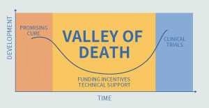
Image Credit: Yokogawa Life Science
Following the requirement for cell-based assay and phenotypic screening, the drug development market is seeing an increase in the demand for High Content Analysis systems for drug efficacy evaluation. Devices with faster speeds (higher throughput) are needed to improve screening efficiency.
On the other hand, better screening hits are needed to cross the “valley of death” in the drug development process.
To achieve this, more intricate assessment methods must be developed, including live-cell imaging, 3D cultivation systems, and higher-detail image processing to leverage numerous aspects.
Determining the best way to conduct parallel throughput and complicated evaluation system screening in drug development research is a crucial problem.
Solution
The high-end High Content Analysis system, CellVoyager CV8000, resolves this contradicting screening problem. They have achieved high throughput, high-resolution imaging as well as phenotypic screening via a more intricate evaluation system by combining a proprietary Yokogawa High-Speed Confocal Scanner, water immersion lens, up to four high field-of-vision cameras, a microscopic stage with cell cultivation environment, and an integrated robotic pipetter.
CellPathfinder, a specialized analysis program, supports users from image analysis to results presented in graphs by utilizing deep learning and machine learning to identify target objects with high accuracy.
The Yokogawa advantage
- Confocal scanner unit
- Live/kinetic experiment compatible
- High throughput
- Reliable, proven technology
Voyage to unknown worlds
Real-time confocal, label-free imaging
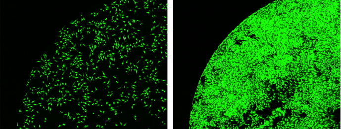
Left: Before incubation. Right: After 68 hours of incubation. Image Credit: Yokogawa Life Science
A stage incubator is provided as usual. Users can implement nonstop, long-duration observation (3 days or more) using humidity, temperature, and CO2 control.
Kinetic assay
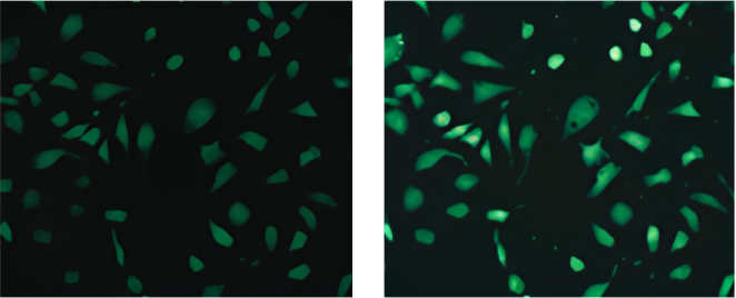
Left: Before incubation. Right: After stimulation. Image Credit: Yokogawa Life Science
An integrated robotic pipette with disposable tips enables drug addition during imaging, making it ideal for high-speed phenomenon monitoring in kinetic experiments.
Organoid/Spheroid
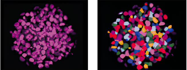
Left: Original image. Right: Recognition image. Image Credit: Yokogawa Life Science
Yokogawa’s spinning disk confocal technology excels in imaging deep samples, such as 3D culture samples, where clear and rapid imaging is difficult. This allows for evaluations that are similar to in-vivo quality.
Label-free analysis
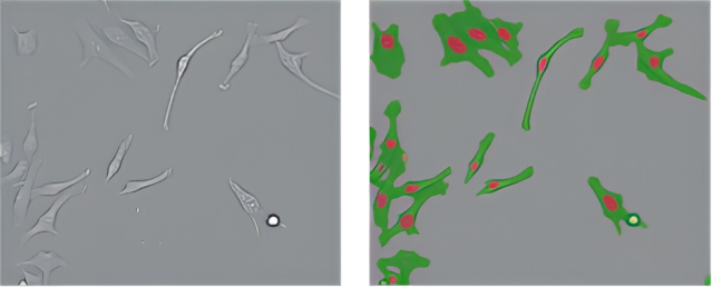
Left: CE Bright Field. Right: Cell Recognition image. Image Credit: Yokogawa Life Science
To perform recognition and analysis, bright field images from various Z locations are converted to CE bright field images using the included CellPathfinder analysis program. The new Deep Learning option substantially improves analysis accuracy.
Details
State-of-the-art technology
State-of-the-art technology that enables users to do what they want
Observe cells as they are - Dual spinning disk confocal system-
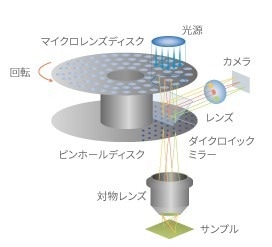
Image Credit: Yokogawa Life Science
Yokogawa's patented multi-scan technology employs roughly 1,000 laser beams on the observation region and tandem disks revolving at high speeds. The disks consist of a pinhole array disk with roughly 20,000 pinholes placed in an equal-pitch spiral pattern and a microlens array disk, which focuses the excitation light laser into individual pinholes. This allows for high-speed imaging and significantly reduces phototoxicity and fluorescence photobleaching.
Deeper, clearer observation -Pinhole disk exchanger-
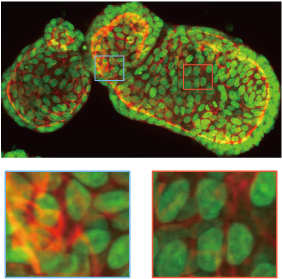
Organoid imaging example. Upper:25 μm pinhole. Lower:50 μm pinhole. Image Credit: Yokogawa Life Science
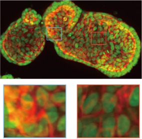
Organoid imaging example. Upper:25 μm pinhole. Lower:50 μm pinhole. Image Credit: Yokogawa Life Science
Depending on the sample, two types of pinhole disks (25/50 μm) can be utilized. For thick materials, lowering the pinhole width increases confocality and shaper images. For dark samples, increasing the pinhole diameter produces brighter images.
Higher throughput screening - Optical configuration-
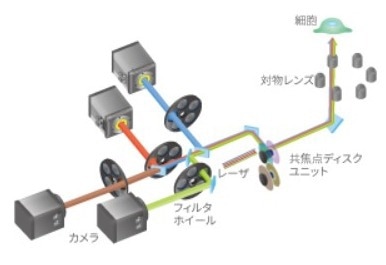
Image Credit: Yokogawa Life Science
The optical system configuration can be chosen based on the purpose. Four high-sensitivity wide-field sCMOS cameras can image a single 96-well plate in four colors in one minute. The system also works with FRET and CellPainting assays.
Capturing finer structures -Original water immersion lens-
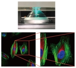
Image Credit: Yokogawa Life Science
High-resolution pictures of liquid-bound cells may be efficiently captured using water immersion lenses. The water immersion objective lens for the CV8000 comes in 40× and 60× magnifications.
The 40× lens is exceptionally unique as it has extremely sophisticated spherical aberration correction, which enables the creation of brilliant, high-resolution images throughout the broad field. The lens water supply is fully automated, making high-throughput screening using a water submersion lens feasible.
Capturing live cell movement - High-precision incubator and robot pipetter
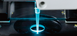
Image Credit: Yokogawa Life Science
The stage incubator's airtight design controls temperature, CO2 levels, and humidity. The integrated robotic pipetter entirely automates the following steps: tip pickup, reagent collection from the reagent plate, reagent addition to the sample plate, and tip disposal.
Reagent instillation allows for the quick acquisition of images both before and after the reagent is added. It also allows for the addition of reagents to a single well several times and the modification of the pace of addition, among other settings, thereby expanding the scope of dynamics observation.
A more live cell-friendly total HCA system
Making long-duration live-cell imaging possible - Featuring a s table built -in stage incubator
500 HeLa cells were planted at a density of one per well on a 96-well plate, and the cells were cultivated for a full day. Following 72 hours of cell culture in the internal stage incubator, the well plate was examined to determine the total area (henceforth referred to as Total Area) occupied by the cells.
Consequently, compared to a standard CO2 incubator, there was barely any variation in cell multiplication throughout the 96 wells (except from the four corner wells).
Comparing the results of 72 hours of incubation with a standard CO2 incubator for cell multiplication.
- 96 well average: 90
- Average of outermost 36 wells: 81
- Average of 60 wells (excl. outermost): 96
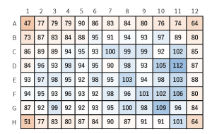
Image Credit: Yokogawa Life Science
The following is represented by the values: CV8000 Total Area after 72 hours/Total Area at 0 hours (hence, Total Area ratio) / CO2 incubator Total Area ratio × 100.
(Numbers near 100 indicate that cell multiplication was almost equal between the CV8000 and the CO2 incubator.)
Cell multiplication was found to be comparable to that of the CO2 incubator, with the exception of the four corner wells.
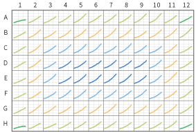
Image Credit: Yokogawa Life Science
Cell multiplication curves for each well in a 96-well plate.
- Vertical axis: Total Area
- Horizontal axis: Time (0-72 hours)
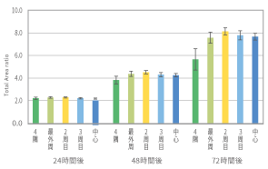
Image Credit: Yokogawa Life Science
Cell multiplication was minimal in the four corner wells but continued in the remaining wells.
Total area ratio after cultivation start (24, 48, and 72 hours) (n = 3).
Excluding the four corner wells, there were no significant changes in cell multiplication after 72 hours.
The minor variance in cell multiplication pace throughout the wells could be observed across 24 hours, 48 hours, and 72 hours.
System integration
Centralized control over all processes, including data management, imaging, analysis, transmission, and cultivation environment. To meet the demands of its clients, Yokogawa provides the best systems.
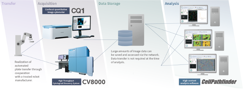
Image Credit: Yokogawa Life Science
High Content Analysis Software CellPathfinder
The software generates graphs, exports several data types, and analyzes image data taken with the CV8000. Due to the software’s numerous templates and adaptable protocol modifying features allow novice and expert users to fully utilize it.
CE’s bright field and machine learning capabilities enable label-free analysis. A new deep-learning option was also included, significantly increasing cell detection accuracy.
Just click the menu item for analysis
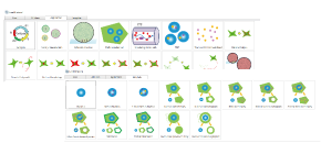
Image Credit: Yokogawa Life Science
Users can follow the flow that appears at the top of the screen. The icons in the analysis menu are straightforward to grasp. They can then choose the relevant menu item, and the protocol will load.
Fast results for immediate verification and study
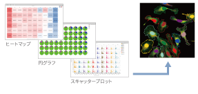
Image Credit: Yokogawa Life Science
Computed numeric data can be shown in a variety of formats. Graph plots and cell images are connected, allowing for simple result verification and inquiry.
Unbiased phenotype evaluation via AI

Image Credit: Yokogawa Life Science
Machine learning also enables the bias-free digitization of visually rated trials. Users can enable automatic recognition by simply selecting the shape they wish the software to learn.
Label-free phenotype analysis
Image Credit: Yokogawa Life Science
Eliminates the time, expense, and impact on cells involved with cell labeling. Combining deep learning allows for even greater accuracy in categorization.
Specifications
High-throughput cytological discovery system
Source: Yokogawa Life Science
| |
|
| Model |
CV8000 |
| Sample format |
Multiple well plate (6, 12, 24, 48, 96, 384, 1536 wells), glass slide |
| Image mode |
Confocal mode: max. 4 color simultaneous recording
Bright-field/phase contrast (10x, 20x for 6, 12, 24 well plates), digital phase contrast (10x, 20x) |
| Output data format |
Image data: 16-bit TIFF, PNG
Numerical data: CSV, original format |
| Excitation wavelength |
405/445/488/561/640 nm, all solid laser, max. 5 lasers
[Option] 365 nm LED |
| White light illumination |
LED |
| Autofocus |
Laser-based mode, image-based mode |
| Objectives |
Max. 6 lenses are available, automatically switchable
Dry: 2x, 4x, 10x, 20x, 40x Water immersion: 20x, 40x, 60x
Phase contrast: 10x, 20x Long working distance: 20x |
| Confocal unit |
Microlens-enhanced wide-view dual Nipkow disk confocal scanner, 50 μm pinhole
[Option] 25 μm pinhole disk and auto pinhole disk exchanger |
| Camera |
sCMOS (effective pixels: 2000X2000 pixel size: 6.5 μm), max. 4 cameras |
| Stage incubator |
Temperature for incubation : 35-40 ℃ CO2 supply box (CO2: 5 %, forced humidification) |
| Robot pipetter |
[Option] Disposable tip type (96 tip or 384 tip type) |
| Bar code reader |
[Option] 1 or 2 dimension |
| Workstations |
Dual-monitor workstation for system control, dual-monitor workstation for data analysis |
| Analysis software |
High Content Analysis Software CellPathfinder
Granularity, Neurite, Nuclear morphology, Nuclear translocation, Plasma membrane translocation, Machine learning, Label-free analysis, 3D analysis, Deep Learning, etc. |
| Operating environment |
15~30 ℃ 30~70 %RH (no condensation) |
| Power supply |
Measurement unit: AC100-240 V, 50/60 Hz, 2 KVA max
Workstation for system control: AC100-240 V, 50/60 Hz, 1.3 KVA max
Workstation for data analysis: AC100-240 V, 50/60 Hz, 950 VA max |
| Dimensions |
Measurement unit: W1,280×D895×H1,450 mm |
| Weight |
Measurement unit: 510 kg
|