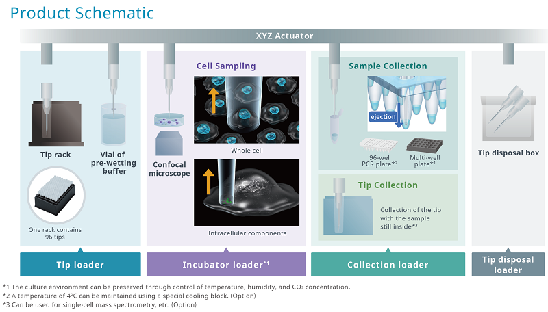
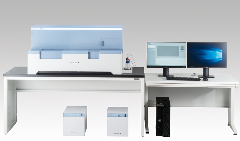
The SS2000 is a dual microlens spinning disk confocal microscope designed for live-cell, high-content imaging. It can sample adherent single cells and subcellular material while preserving spatial, morphological, and temporal data.
Single-cell and subcellular sampling process

Image Credit: Yokogawa Life Science
- Automated, intuitive operation improves productivity and efficiency
- Image-based localization ensures precise cellular sampling
- Sampling preserves information about morphology and space
- Confocal microscopy for high-resolution live cell imaging and image analysis
- Integrated incubator enables live cell experiments
- Adaptable environmental control to preserve the integrity of samples that have been collected
Product specifications

Image Credit: Yokogawa Life Science
Source: Yokogawa Life Science
| |
|
|
| Automatic sampling functions |
Tip diameter |
3 μm, 5 μm, 8 μm, 10 μm |
| Incubator loader environment |
37 ℃, 5 % CO2, humidified |
| Collection loader environment |
37 ℃, 5 % CO2, humidified (for culture) / 4 ℃ (for cooling) |
| Collection loader-compatible vessels |
96-well PCR plate (0.1 mL, 0.2 mL)
Multiwell culture plate (96 well) |
| Postioning precision of sampling |
XYZ axial designated resolution: 0.1 μm |
| Imaging functions |
Confocal scanning method |
Microlens enhanced dual wide Nipkow disk confocal |
| Incubator loader-compatible vessels |
When sampling cell: φ35 mm dishes *1
Microplate (6 well, 24 well, 96 well) |
When observing cell: φ35 mm dishes *1
Microplate (6 well, 12 well, 24 well, 48 well, 96 well, 384 well, 1536 well)
Slideglass *2 |
| Excitation laser wavelength |
405, 488, 561, 640 nm (Uniformizer installed) |
| Emission filter |
Filter size: φ25 nm
Maximum slot number: 10 (Electric switching)
Adjacent switching speed: 100 msec |
| Transmission illumination |
Bright-field, LED source |
| Objective lens |
Dry lens: 4x, 10x, 20x, 40x
Long-working distance lens: 20x, 40x
Note that only the 40x dry lens can be used for cell sampling. |
| Z focus |
Electric Z motor, designated resolution: 0.1 μm |
| Electric stage |
XYZ axial designated resolution: 0.1 μm |
| Autofocus |
Laser autofocus |
| Camera |
sCMOS camera 2,000 x 2,000 pixel Pixel size:6.5 x 6.5 μm |
| Other |
Special purpose workstation |
Workstation for sampling, measurement, analysis, 24-inch display x2 |
| Measurement software |
Measurement functions (2D, 3D, Time-lapse, Map imaging), Viewing measurement and sampling data, Reporting functions (Image data, Video data), Whole-cell sampling, Intracellular component sampling |
| Analysis software |
Analysis functions (3D, Tile, Label-free, Texture analysis, Deep Learning, Gating), 3D viewer, Graphing functions, Reporting functions(Image data, Video data, EC50, IC50, Z'-factor) |
| External dimensions, Weight |
Main unit: W1,217 x D643 x H595 mm, 145 kg
Utility box: W275 x D432 x H298 mm, 18 kg
Gas mixer: W275 x D432 x H298 mm, 10 kg
Special purpose workstation: W172 x D471 x H414 mm, 14 kg
Display: W531 x D500 x H166 mm, 5.6 kg |
| Operating environment |
Temperature: 15 to 30 ℃
Humidity: 30 to 70 %RH no condensation |
| Power consumption |
Main unit, Utility box and Gas mixer: 1,200 VAmax
Workstation: 950 VAmax
Display: 42 VAmax x 2 |
| Data formats (Measurement software) |
Captured images: 16-bit TIFF (OME-TIFF, TIFF)
Output image data: TIFF, PNG, JPEG
Output video data: WMV, MPEG4 |
| Data formats (Analysis software) |
Numeric data: CSV
Output image data: TIFF, PNG, JPEG
Output video data: WMV, MPEG4 |
*1 A sample holder is required, and with it, up to 3 samples can be installed.
*2 A sample holder is required, and with it, up to 4 samples can be installed.