The CellVoyager High-Content Analysis System CQ3000 can quickly and efficiently capture high-resolution 3D images when culturing cells.
CQ3000 can be configured with choices based on the intended usage to create a system that meets the desired goals.
Make way for high-performance HCA optimized for laboratories
Depending on the application, a second camera for simultaneous imaging of two channels, a high NA immersion lens for high-quality imaging, and a uniformizer for uniform and efficient imaging of the full FOV can be chosen.
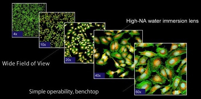
Image Credit: Yokogawa Life Science
High-resolution images that hold tons of information
- Water immersion lenses produce clear images with a high SNR
- In addition to precise observation via high magnification, the high-NA lens allows for reliable image analysis even with lower FOVs than before
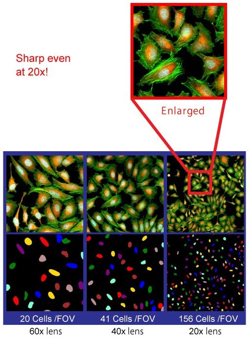
Image Credit: Yokogawa Life Science
Developed in-house, the high-NA 20× water immersion lens achieves greater image resolutions than a dry lens. Users can analyze images more efficiently by using 40× and 60×.
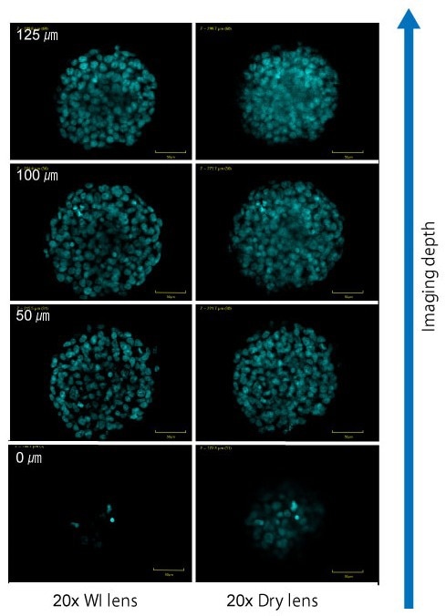
Image Credit: Yokogawa Life Science
- Imaging example of a spheroid
- The 20× WI lens enables deeper imaging with high SNR
High throughput imaging via simple operation
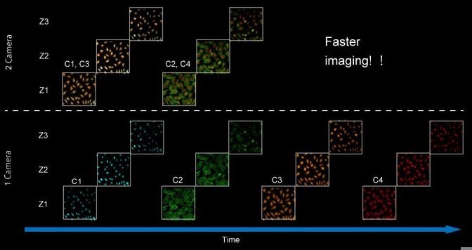
Image Credit: Yokogawa Life Science
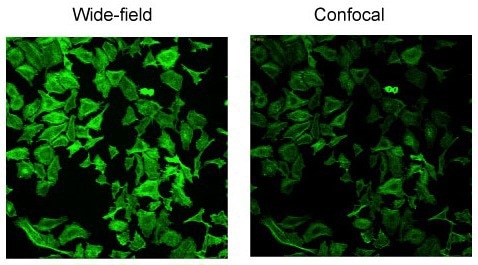
Image Credit: Yokogawa Life Science
- Comparing throughput with the second camera option
- Detects two colors simultaneously, reducing acquisition time
- Furthermore, widefield imaging can further reduce the exposure time if confocal performance is not required, as in the case of low-magnification imaging
When it comes to Yokogawa, it is all about live-cell imaging
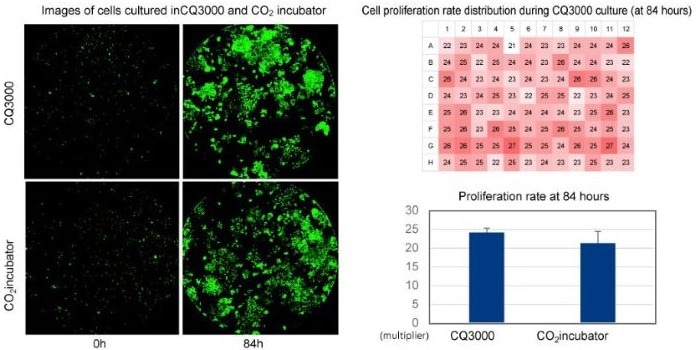
Image Credit: Yokogawa Life Science
With CQ3000, users can image and culture in the same atmosphere as a conventional CO2 incubator.
It reduces the effect on cells and is also good for the environment.
Details
CellPathfinder
Analysis software: High content analysis system CellPathfinder (option)
The clear, easy-to-use interface walks the user through the process of evaluating hundreds of images from various perspectives and visualizing the results using a variety of graph styles.
In addition, the Machine Learning and Deep Learning functions significantly improve target recognition capabilities. It is also suited for sophisticated and demanding analyses, such as those involving 3D culture and live-cell imaging.
For HCA, the CellPathfinder software is an effective tool.
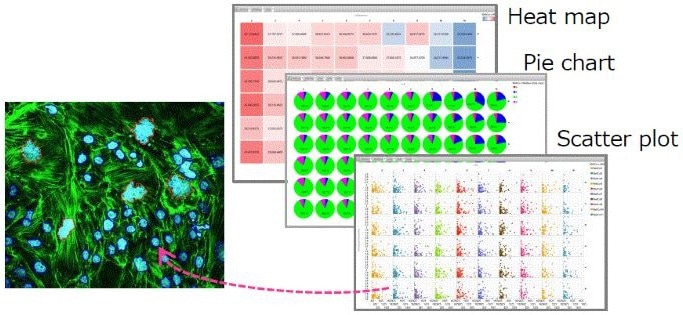
Image Credit: Yokogawa Life Science
Machine Learning
The software picks up characteristics from sample objects that users have gathered.
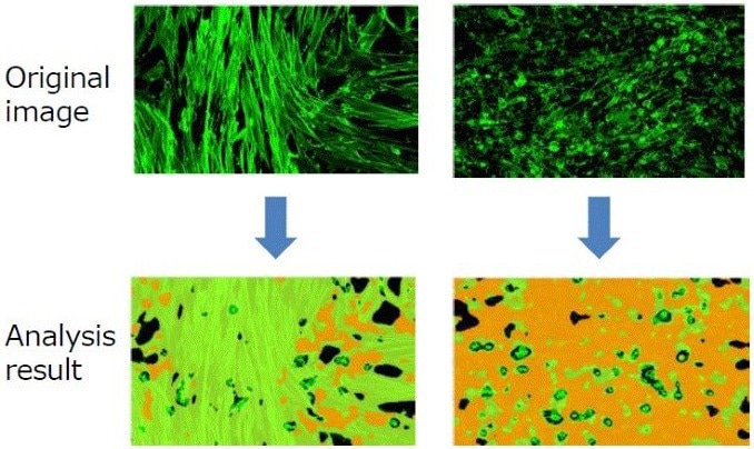
Image Credit: Yokogawa Life Science
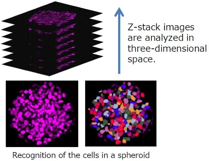
Image Credit: Yokogawa Life Science
Label-free analysis
The wide-field function is an effective tool for analyzing unstained bright-field samples.
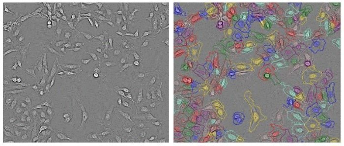
Image Credit: Yokogawa Life Science
Specifications
Source: Yokogawa Life Science
| |
|
| Optics |
Microlens enhanced dual wide Nipkow disk confocal, Transmitted illumination |
| Fluorescence |
Laser: Up to 4 colors Standard: 488 nm, 561 nm Option: 405 nm, 640 nm
High-power lasers option: 488 nm, 561 nm
EM filter: Max. 10 filters (including 1 filter for transillumination)
Observation method: Confocal image, Wide-field image*1 |
| Transmitted illumination |
Bright-field LED light |
| Camera |
High-sensitivity sCMOS
Max. 2 units, simultaneous excitation of 2 wavelengths
Number of effective pixels: 2000 x 2000 pixels
Field of view size: 13.0 x 13.0 mm |
| Objective lens |
Max. 6 lenses (Included max. 2 water immersion lenses)
Dry: 2x, 4x, 10x, 20x, 40x, 60x
Long working distance: 20x, 40x
Water immersion: 20x, 40x, 60x |
| Water supply function for immersion lens |
Automatic supply |
| Flat-top beam shaper Option) |
Uniformizer |
| Sample vessel |
Microplate (6, 12, 24, 48, 96, 384, 1536 wells), glass slides*2, cover glass chamber*2, 35 mm dish*2 |
| Stage incubator |
Temperature control range: 35 ~ 39 °C
Settable temperature resolution: 0.1 °C
Time stability: ±0.2 °C
Spatial stability :±1 °C (at an ambient temperature of 21 ~ 25 °C)
Humidity holding
Automatic water supply function for incubator |
| Autofocus |
Laser autofocus, Image-based autofocus |
| Fast time-lapse (Option) |
Max.100 fps, Simultaneous dual-wavelength excitation imaging |
| Analysis software CellPathfinder) |
Granule analysis, Neurite analysis, Nuclear morphology analysis, Nuclear translocation analysis, Membrane translocation analysis, Machine learning, Label-free analysis, 3D analysis, Texture analysis, Deep Learning, etc. |
| Other features |
Self-diagnosis function, CQ Analysis |
| Size・Weight |
Main unit W1031 mm x D401 mm x H600 m 84 kg
Main unit (with Uniformizer or 2nd camera) W1177 mm x D401 mm x H600 mm 102 kg
Utility box W275 mm x D432 mm x H298 mm 17.6 kg
Gas mixer W275 mm x D432 mm x H298 mm 9.3 kg
Workstation W176.5 mm x D452.1 mm x H417.9 mm 21.7 kg |
| Operating environment |
15 ~ 30 °C, 30 ~ 70% RH, no condensation |
| Power consumption |
Main unit and Utility box: 100 - 240 VAC, 400 VAMax
Gas Mixer : 100 - 240 VAC, 60 VAMax
Workstation: 100 - 240 VAC, 750 VAMax |
*1 Can be acquired when Uniformizer option is selected.
*2 Sample Holder option is needed.