An interview with Samuel Lesko, European Applications Manager, Bruker Nano Surfaces conducted by April Cashin-Garbutt, MA (Cantab).
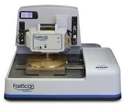
Can you please give an overview of the FastScan AFM system?
The FastScan offers the ability to scan up to 4 frames per seconds. We can interact live with the system, pan & zoom in real time, and see nanometer details that could evolve over time. Once we collect all the data, we can generate automatically a movie, which can be a movie offline or real time as the data is added one by one.
What applications has the FastScan been designed to address?
Regarding application, the Bio FastScan has been really designed to address a specific segment of the AFM applied to the biological materials.
First of all, we like to study dynamic processes - it can be from lipid bilayers, macromolecules or proteins interactions, and really map topography as well as real-time mechanical properties while we are measuring.
This leads us to an understanding of what processes are going on, such as membrane formations or amyloid fibril growth in a way that we can, by better understanding, address disease and test drugs.
With the FastScan, we mainly address biomolecules, proteins, aggregations, as well as bacteria, and some further extend up to living cells in respect to what we scan around.
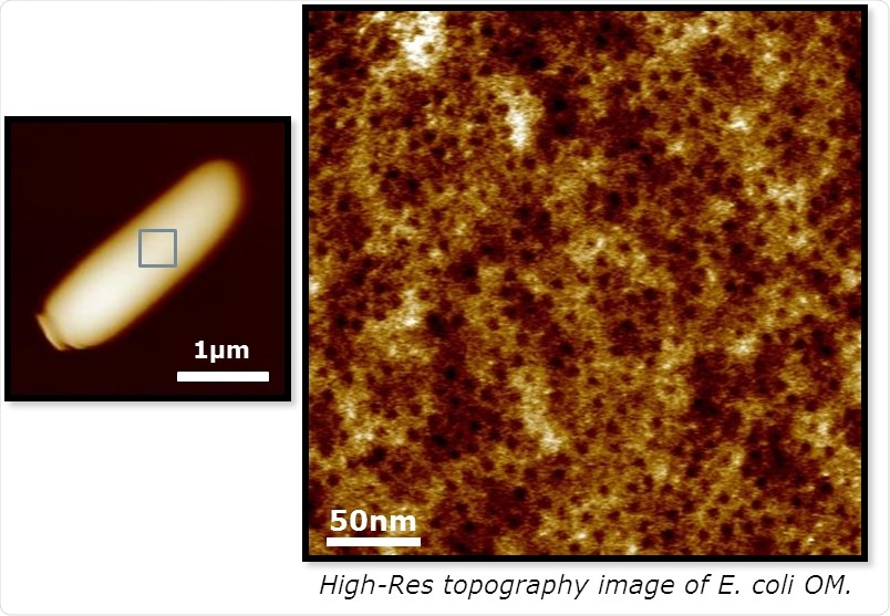
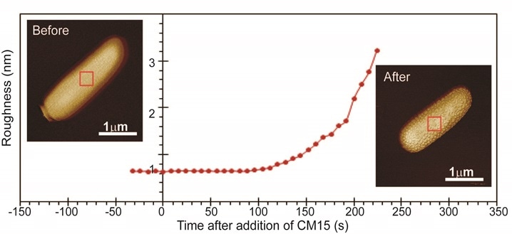
Top: high lateral resolution on living bacteria in saline buffer solution. Bottom: time lapse resolved action of peptide disrupting bacteria membrane
How important is the ability to scan fast?
In biology there are many samples, which can differ one by one, so, at some point you need to speed up the whole imaging process in a way you collect statistics. It is not the question of scanning fast for scanning fast, it is a question of collecting enough data to draw a conclusion and the accuracy of that conclusion will depend on how much sample we've processed.
The idea of the FastScan is to create a platform where you can place multiple samples and automatically target and measure them without the user attending the system.
We designed the modes like with the ScanAsyst that will automatically adjust scan and set point with respect to conditions, as well as design a track where you can just travel to different wells, for instance, and measure living cells or measuring mechanical properties. And then the software offline will help you to process this data in the batch in order to create a database and then draw accurate conclusions.
What changes have you seen in ease of use?
Previously, users had to spend hours finding the right spot and now with the FastScan we simply zoom in, zoom out, engage fast and quickly move to different locations. We can even automatically probe this location without the user wasting his or her time.
Can you tell us about any case studies that demonstrate the FastScan systems ability?
Long term stability of DNA double helix structure: AFM performances of Dimension Fastscan combined with high lateral resolution capability provided by Peak Force Tapping ensured seamless operation at such high lateral resolution. It opens up further studies toward triplex formation as well as binding of protein on DNA.
A second example looks directly at thevisualization of peptide action to disrupt bacteria membranes, which provides a new mechanism of action for a new range of drugs against resistant bacteria over common antibiotics.
A third example concerns fibril formation such amyloids. Kinetic study as well as formation mechanism are key to better understand fibril formation and develop strategies to efficiently prevent it or fight its extension.
Overcoming longstanding barriers associated with imaging live cell interactions is a huge step forward for this technology. How do you think this will benefit cell dynamic research?
Dimension Fastscan efficiently provides information on how stress fibres and fibrils from cytoskeleton re-organize versus external stress or during cell motion. It brings new information that helps understand tumour spread as well as the recovery of tissue from injury. Last but not least, Dimension Fastscan enables direct or indirect visualization of pathogen/host cell interactions through topography or receptor mapping with functionalized probes.
Where can readers find more information?
About Samuel Lesko
Samuel Lesko has more than 20 years experiences in running AFM over variety of applications.
After completing a PhD at Burgundy university on colloidal force measurement in cement, he started his career at Veeco Instruments as French Applications Scientists before to continue on supporting Bio Applications AFM European wide.
He is since 2007, Applications manager for Europe, Middle East and Africa. Recently, his position expanded to Latin America.
About Bruker Nano Surfaces and Metrology

Bruker’s suite of fluorescence microscopy systems provides a full range of solutions for life science researchers. Their multiphoton imaging systems provide the imaging depth, speed and resolution required for intravital imaging applications, and their confocal systems enable cell biologists to study function and structure using live-cell imaging at speeds and durations previously not possible. Bruker’s super-resolution microscopes are setting new standards with quantitative single molecule localization that allows for the direct investigation of the molecular positions and distribution of proteins within the cellular environment. And their Luxendo light-sheet microscopes, are revolutionizing long-term studies in developmental biology and investigation of dynamic processes in cell culture and small animal models.