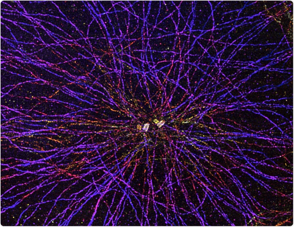
Researchers at the University of Geneva have developed a technique that enables visualization of cellular organelles at a resolution that has not previously been achievable in optical microscopy.
 UNIGE
UNIGE
Being able to see these structures would allow an improved understanding of how cells function, but achieving this has been a difficult challenge. Until now, fluorescence microscopy has not provided a resolution high enough to observe the ultrastructure of these components.
Now, University of Geneva researchers have developed a technique that enlarges biological samples without destroying or deforming them and observe them at a nanometric scale – an unprecedented resolution in optical microscopy.
As reported in the journal Nature Methods, the method enables visualization of the composition and architecture of organelles and protein complexes.
It even enables detection of biochemical modifications on components of the complexes, which could be useful for cellular mapping.
Study author Professor Paul Guichard says that it all began three years ago when Professor Edouard Boyden from the Massachusetts Institute of Technology developed of a way of embedding cell structures with a mixture of sodium acrylate and acrylamide.
Next, he marked target structures with fluorescent molecules before swelling the sample by adding water.
The targets had to be destroyed, but it was possible to visualize their fluorescent borders with a good resolution, thanks to the expansion obtained.”
Professor Paul Guichard, Lead Author
Co-author Virginie Hamel says the team wanted to find out whether the technique could be adapted to observe organelles without destroying them and enlarge them without deforming them.
Eventually, the team developed a method which enables a biological sample to be inflated while maintaining its native state and without the use of chemicals that would denature it.
Cells gradually expand, and their components separate from each other while enlarging. The architecture of the various elements is preserved, and it becomes possible to observe them with a resolution hitherto unattained in optical microscopy.”
Davide Gambarotto, First Author
The technique, which is called Ultrastructure Expansion Microscopy (U-ExM), enables nanoscale visualization of cellular structures that had previously only been possible using electron microscopy. However, electron microscopy does not enable proteins that make up the cellular elements to be located.
“Our method combines the advantage of fluorescence microscopy to detect molecules and high resolution to visualize the fine structure of organelles or of macromolecules," explains Hamel.
"It is now becoming possible to map large intracellular molecular complexes. This method could also be used to reveal signatures of pathological processes at the very heart of the cell," concludes Guichard.