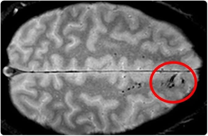
Microbleeds are small areas of hemorrhage that are not detectable by CT scanning but are picked up by MRI as tiny dark patches. They are caused by blood vessel injury, and may be found after apparently even minor degrees of head injury. The problems caused by this type of damage to blood vessels is not quite clear but since any injury to brain cells can be potentially wide in its impact, the researchers feel that microbleeds need to be studied in greater depth.

Traumatic microbleeds appear as dark lesions on MRI scans and suggest damage to brain blood vessels after head injury. Image Credit: Latour Lab/NINDS.
CT vs MRI scanning
A computerized tomography scan (CT scan) of the brain is performed with the help of X-ray machines which emit low-energy X-rays while rotating around the patient’s body, focusing on the part to be imaged. The images are sent to a computer which collates images of the same plane taken from different angles as the scanner moves around the body, to create a detailed image, including soft tissues, blood vessels and bones unlike a standard X-ray. The image appears as a virtual cross-section taken through the part to be imaged, thus providing a view inside the body without actually cutting it open. At present, CT scanning is the most frequently used form of imaging when doctors need to quickly visualize possible internal injuries following trauma such as vehicular accidents.
An magnetic resonance imaging (MRI) scan is a medical imaging technique that uses an applied strong magnetic field to force the body’s protons to line up with it. A stream of radiofrequency waves is then passed through the body, which excites the aligned protons to spin wildly, against the applied field. After the RF current is switched off, the extent of shift in the protons as they return to their original forced alignment is sensed by the amount of energy they release, by special energy sensors. The period taken for realignment and the amount of energy released change with the type of environment and the molecular chemistry, which has served as the basis for physicists to interpret with confidence what internal tissues are being imaged, and the kind of changes being observed. MRI scans show soft tissues very well, but avoid the use of ionizing radiation like X-rays – two advantages this technology has over CT scanning.
The study and its findings
The current study was carried out on 439 adults with head injury who were seen in the emergency department. All of them had MRI scanning within 48 hours of the trauma and the scans were repeated at each of four visits following this initial screening. At the same time they were also asked about behavioral patterns and other neurologic outcomes, using a standard questionnaire.
The findings showed that microbleeds were present on the MRI scan in 31% of all the patients, but the frequency varied with the severity of head injury. For instance, when severe head injury was present, almost 60% of the patients had microbleeds, in contrast with only 27% in mild injury.
The appearance of the microbleeds was also variable – some were linear, while others were punctate (like small dots), and most patients showed both types. The area of the brain most likely to show microbleeds was the frontal region of both hemispheres.
With respect to the outcome, the researchers found that if a patient had microbleeds, the outcome was likely to be associated with greater disability, as assessed by a scale that is widely used.
Confirming MRI findings by physical examination
The researchers were able to confirm their findings and extend them by examining the brain of one participant (donated by the family) who died after the study ended, by still more powerful MRI scanning and histopathological examination showed a sharper level of detail. The tissue analysis showed the presence of iron in the macrophages of the brain along the blood vessel path, and also beyond this in areas that could not be seen with the scan.
Researcher Alison Griffin says, “Combining these technologies and methods allowed us to get a much more detailed look at microbleed structure and get a better sense of just how extensive they are.” This knowledge could help in deciding if post-traumatic microbleeds can be used as a biomarker to identify patients who can benefit from therapy that targets vascular injury.
So should all head injury patients receive an MRI scan rather than the less expensive CT scans? At present the evidence does not support such a shift. Deeper analysis will be required to find out if there are other effects caused by microbleeds, as well as therapies to avoid detrimental outcomes. This would also point the direction for future studies to help select the individual patient for whom MRI rather than CT scanning is indicated.
Journal reference:
Griffin, A.D., et al. (2019) Traumatic microbleeds suggest vascular injury and predict disability in traumatic brain injury. Brain. doi.org/10.1093/brain/awz290. https://academic.oup.com/brain/advance-article-abstract/doi/10.1093/brain/awz290/5581280?redirectedFrom=fulltext