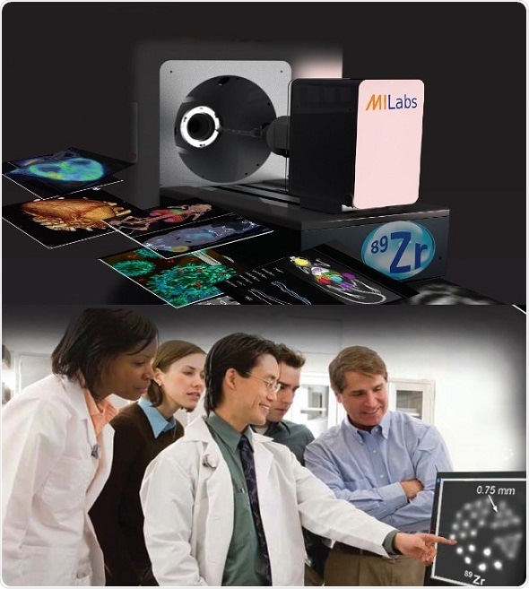
Researchers at MILabs B.V. have succeeded at imaging the popular Zirconium-89 (89Zr) isotope in small animals of human disease at very high resolutions (<0.75 mm). 89Zr as used for Positron Emission Tomography (PET) has great appeal to immune-oncology researchers because it is a nearly ideal radioisotope for PET imaging with immunoconjugates, as it possesses a multi-day physical half-life that is compatible with the in vivo pharmacokinetics of antibodies. Unfortunately, its positron-rage of 1.3-3.9mm and background of high energy prompt gamma’s causes non-isotropic blurring and haziness in the PET images of small laboratory animals making quantification and mapping of tumor heterogeneity in mouse oncology models virtually impossible. Quantification of tracer uptake and accurate assessment of tumor heterogeneity is considered by cancer researchers as essential for the development of effective therapies.

By using information of prompt photon emissions and advanced modeling of high energy photon transport during image reconstruction, the researchers at MILabshave succeeded at dramatically improving the three-dimensional imaging precision over conventional PET. Moreover, since MILabs’ PET systems can image multiple isotopes, 89Zr-immunoPET therapies and 18F-FDG cancer staging can now be performed simultaneously in the same animal at high-resolution to augment cancer therapy research.
Prof. Frederik Beekman, CEO/CSO MILabs states:
We are exited that we have been able to complete our mission of making imaging clear for difficult PET isotopes by eliminating the positron range blurring effect from small animal imaging for three important PET isotopes: 82Rb for cardiac imaging, 124I for e.g. oncolytic virus therapy imaging and now 89Zr for immunotherapy imaging.”