An interview with Mark White, Ph.D., discussing the need for single-cell analysis and techniques that allow the isolation of high-value T-cells for use in academic research and pharmaceutical development.
Why is the pharmaceutical industry interested in identifying the function and phenotype of individual T-cells?
Over the past decade, there has been an explosion in the development of immunotherapies; drugs that turn the immune system into a weapon against cancer or another disease state. With the emergence of immuno-oncology-based antibodies and cell therapies, there's a huge interest in T-cells.
Pharmaceutical companies want to know what the T-cells are doing, how they function, and how we can exploit this for the creation of better therapeutics. This underlying interest has translated into looking at T-cells in a whole new way and diving deep on the phenotype and function of T-cells.
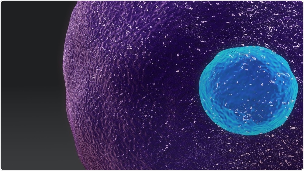
 Image Credit: sciencepics / Shutterstock.com
Immunologists have been looking at individual T-cells for a long time. The goal of the pharmaceutical industry is to find the best cells. If you're making a cell therapy and you're putting billions of cells into a patient to try and treat their condition, one of the key questions is, how many of those cells have the function that you think they do? The function being quite complicated cell behavior, which is the ability to interact with a tumor cell, recognize it as foreign and then attack it.
Most of the questions surround two key themes; how can we make our processes more efficient and how can we identify only the best cells? Scientists working in this field are looking at ways to identify and link directly with the phenotype of an individual T-cell with its actual cell behavior or function.
Following this, in an R&D setting, the scientists will try to make T-cells more functional, looking at the genomics or the underlying transcriptome of a T-cell and asking why one group of T-cells was able to kill a group of tumor cells and another wasn’t.
If you are able to deeply characterize, profile and map your T-cell populations at the single cell-level, you can start to identify the fraction of T-cells that are actually doing what you want.
What techniques are typically used for this process in the pharmaceutical industry? Are there any limitations?
There are a couple of key limitations for the common techniques that are done today. The first relates to high-throughput techniques that only allow you to take a single snapshot of that population in time.
You may be able to achieve single-cell resolution of what each of those individual T-cells looks like at that timepoint, but if you then change something or continue to culture, you cannot directly link that cell's phenotype or behavior at the first time point to the second.
For example, you are unable to link phenotype for a single cell a timepoint 0 with gene expression or the cells’ phenotype at the 12-hour timepoint. This prevents you from getting the highest resolution data on the best cell behavior or genotype for a successful therapy.
Another issue comes in with live-cell imaging or methods that can test individual T-cell function. With most of these techniques, you can't recover the cells from the device or instrument that you're taking the measurement from.
At Berkeley Lights, our platforms can be used to track thousands of single T-cells over time, taking multiple measurements of cell behavior or phenotype and then finally looking at their genotype. Through this process, scientists can find the best cells within the parameters they are looking for.
The ability to kill a tumor cell combined with Alpha-beta chain sequences from a single T-cell, for example, will define ways to target T-cells towards a specific antigen. Scientists may look at the whole transcriptome to try and ascertain what was happening in that T-cell and why that differed to another cell from the same population.
The third and final limitation is the destruction of precious samples. All human samples are precious, and most of them come in relatively small volumes with limited numbers of cells. It is therefore vital that you can recover most or all of the sample after analysis. To achieve this, scientists working in the field of immunology are always looking for ways to integrate measurements together into the same chip, on the same cells and get as much information as possible.
For scientists carrying out cell therapy manufacturing, they must first analyze their process using samples. If it’s a real patient sample, they can’t afford to lose many of those cells while trying to understand or optimize the process. Using as few cells as possible and extracting as much information as possible from those cells is valuable to them.
That’s why we developed our Lightning™ and Beacon® platforms, which allow scientists to work with small cell numbers; between 1000 to 100,000 cells in total.
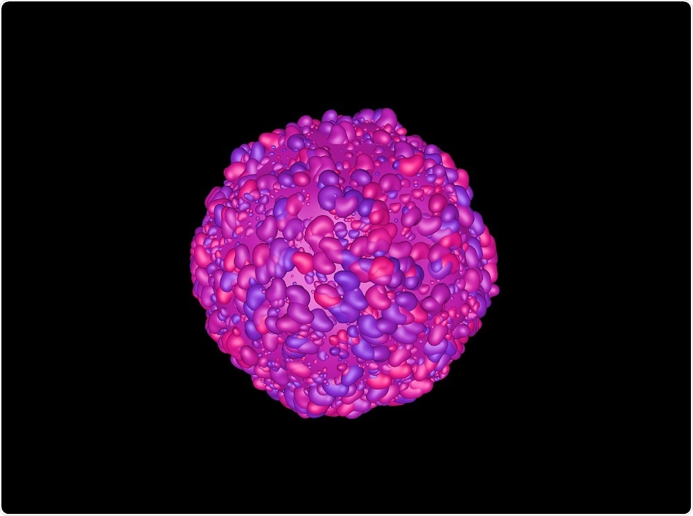
 Image Credit: CI Photos / Shutterstock.com
What are the Beacon and Lightning platforms and how do they work?
We launched the Lightning platform in June. This platform was designed for focused efforts and assay development on T-cells in particular. It has been optimized for immunologists that work in low-throughput laboratories who need to tweak and change their assays often.
They want to be able to quickly change an assay and develop a new one. For them, the automation is not important. What's most important is getting their sample into the system as quickly as possible and carrying out their experiment.
Compared to our Beacon platform, which has been designed for high-throughput applications, the Lightning platform has a more accessible price point. It runs a single OptoSelect chip and has 1,500 nanopens on it. We're able to screen roughly 1,000 single T-cells in a single run over the course of 24 hours.
The Beacon platform was launched about three years ago and has been designed for Cell Line Development and plasma B-cell discovery.
For Cell Line Development, the Beacon platform enables scientists to identify CHO cells that secrete the highest titers of IgG molecules with 99.9% monoclonality in five days. It allows very rapid selection of the best clones.
For scientists working on plasma B-cell discovery, they are carrying out lots of high-throughput screening to find the best lead molecules. The Beacon has been developed to screen 25 to 30,000 plasma B cells directly, without fusion to a myeloma cell to make a hybridoma. These cells are fragile and don’t live very long, so being able to look at each individual cell and quantify, for example, whether the antibody that they're secreting will bind to the antigen on the surface of a live cell, is useful. This process normally takes 3 months, but the Beacon reduces this down to 1-2 days.
How does the Lightning system help R&D scientists to recover T-cells of interest? How do you manage to keep the cells alive during this process?
Any manipulation you do to a single cell carries a risk for that cell. We started the company with the mindset that we want to provide the world’s best environment for a single cell. There are a lot of aspects to this and it’s our OptoSelect chip that underlies everything we develop. We made sure that every surface was highly defined with surface chemistry and that it was highly biocompatible with T-cells, B-cells, or CHO cells.
Each cell is placed into one of our nanopens, so called because they have nanoliter volumes. A single cell in a nanoliter is the equivalent of 1 million cells per mL, which is the ‘happy density’ for most cell types and cell cultures. The cells are able to secrete factors that keep them alive and functioning.
The small volume allows them to create a microenvironment within minutes to an hour that’s highly conducive to cell growth. We also perfuse media through the chip, meaning the cells are constantly receiving fresh media and waste products can be removed by diffusion.
You can think of it like a microcapillary system in the human body. Instead of a static culture where the cells can build up a lot of waste products that make them unhappy, we’re constantly providing media with the right pH, and nutrients and salt concentrations, at the optimal temperature. All of this is fully defined by our users, including which media they are using.
When scientists then start doing imaging, or staining, the cells are in a nice, low impact environment. This enables scientists to do more longitudinal studies that would otherwise not be possible.
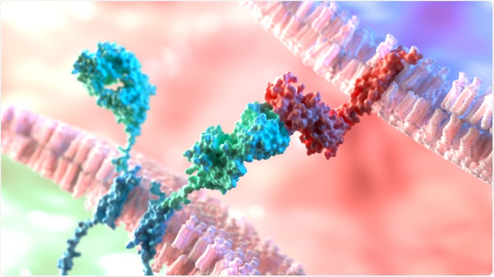
 Image Credit: Alpha Tauri 3D Graphics / Shutterstock.com
Was it important for you to develop a platform that allows scientists to build their own assays?
In the life science industry, we commonly see scientists purchasing a single box that does one specific assay or workflow, and that’s it. There's really very little you can change about it and you either want to do that one workload or you don't.
When we built the Beacon Platform, and then later, the Lightning Platform, the goal was to make it user-friendly and allow our customers to take the basic workflows that we had developed and run them straight away.
If they needed something different, they could modify that workflow and customize it for their own process to make it world-class. We're trying to balance those two with the Beacon, but the Lightning was developed to be flexible with no need for coding experience.
I'm a biologist. I was trained as a molecular biologist and I don't know how to code in Python or how to customize a computer program, but I want to be able to customize the platform that I’m using. For example, I want to be able to easily change when the assay is done, when the cells come in, or how they come in.
We recently did a big overhaul of our software to enable scientists to drag and drop block of modules that allow a biologist to build a workflow or modify workflow that already exists, fairly quickly and easily.
Most of our biologists using the Lightning Platform are up and running within 3-4 days of training. At the moment, we think that's unique for our platform, particularly in an industry where the focus is on developing novel biotherapeutics.
You recently launched the Culture Station, an instrument designed to increase the workflow capacity of the Beacon and Lightning platforms. How does the ‘station’ work, and why did you decide to develop this instrument?
We always ask our customers for feedback, and while most of it was positive for the Beacon and Lightning platforms, some scientists wanted to increase the throughput.
A lot of the questions we got were, "If I load four chips on a Beacon or a single chip on a Lightning, then that experiment has to complete before I start my next experiment. That limits me in some of the throughput that I can get from the platform. Is there a way I could take the chip off, if I just need to grow cells for two to three days and I don't need to image them, and simply grow them? Can I grow them in an incubator?"
Due to the way that the chips work, we need to have a pump that's constantly pushing media through, in addition to maintaining the temperature. You can't just take the chip out like a well plate and drop it in an incubator and maintain the same level of cell viability.
We received this feedback and realized that we had already created the solution internally. Our scientists were using a unique version of the platforms to increase their own throughput. We have a close relationship with our customers, so they knew about this system which was only being used for internal development. They asked if we could provide a product-version of this station, so we did.
Essentially, the accessory allows you to move the chips from a Beacon or Lightning onto the Culture Station, and culture them. It maintains the temperature of the chip and keeps the profusion of media through it during the whole run.
There is no imaging, so we were able to develop a platform that can support up to four chips at the same time. This allows experiments to be run in an increased scale. You could load four chips on a Beacon or a chip on a Lightning and move them off to the Culture Station, then load another chip and so on.
You can double the capacity of a Beacon with a single Culture Station or quadruple the capacity of a Lightning. The other unique feature of the Culture Station is that every chip is independent from a media perspective. You could run four different medias on the same cells across four different chips and look at the effects on cell viability and growth, for example, allowing for media optimization. It also frees up each instrument to start a new experiment.
This is particularly useful for the Lightning platform which is often being used into a shared facility where different researchers want to run multiple experiments each week.
What’s next for the company?
The theme is to provide the most value for our customers. This year will be a big year for us. We are releasing a lot of new products and have already announced the second version of our plasma B-cell discovery platform a couple of weeks ago, followed by the Culture Station.
We are now looking to expand into new applications that we haven’t covered before or updates to existing applications that drive them to the next level, either in the number of cells that are screened or the types of assays that can be done on a single cell.
We just announced a large project with Ginkgo Bioworks in the synthetic biology space. We have grand aspirations to be a large company that provides solutions across a wide array of the life science industry, not just mammalian cells, but also other types of cells. The goal of the company is to allow our customers to find the best cells across the board.
Where can our readers find more information?
About Mark White, Ph.D.
 Mark has been with Berkeley Lights almost from the beginning. He was the first molecular biologist that the company hired to do assay development on their proprietary single-cell secretion assay.
Since then, he has built a genomics team followed by a move into a marketing role around three years ago.
He is currently the Senior Director of Marketing for T-cell discovery. His career highlights include seeing the company grow from 13 people to over 200 and deliver multiple products to market.
We have taken our products from a PowerPoint presentation at the beginning to a real product that customers are using every day. That hasn’t been an easy process because there's engineering, software, surface chemistry, biology, process engineering… all of these things that all had to come together to build the platforms and make them work smoothly for our customers. However, it’s been incredibly rewarding to be a part of the process from start to finish and build a company at the same time," commented Mark.