The spike (S) protein of severe acute respiratory syndrome coronavirus 2 (SARS-CoV-2) binds to the human angiotensin-converting enzyme 2 (hACE2 or ACE2) receptor through the receptor-binding domain (RBD) on the host's cell surface and initiates SARS-CoV-2 infection. Studying spike-ACE2 interactions, particularly the role of glycosylated sites of these proteins, is critical for developing more efficient tests and therapeutics against SARS-CoV-2.
The SARS-CoV-2 S protein is a trimer, where each of its monomers is highly glycosylated. In past research studies, researchers could observe only 16 N-linked glycosylation sites in a cryo-EM map of S protein using the cryogenic electron microscopy (Cryo-EM) technique.
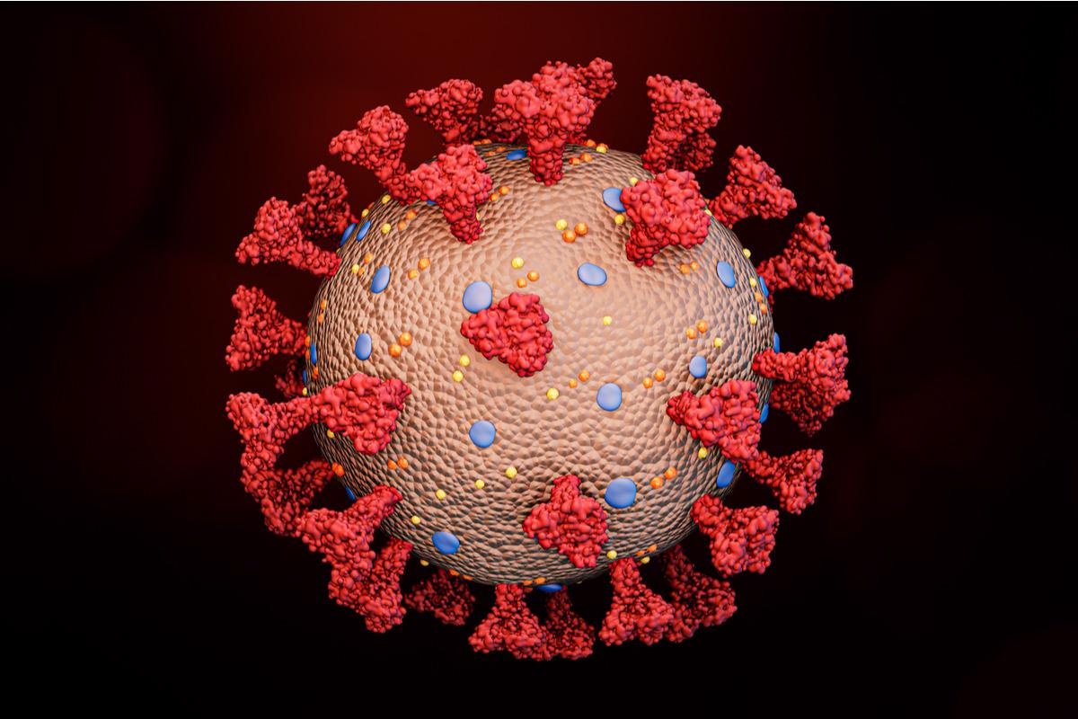
 Study: SARS-Cov-2 Spike binding to ACE2 is stronger and longer ranged due to glycan interaction. Image Credit: MattLphotography/Shutterstock
About the study
A recent study posted in the Biophysical Journal addressed this gap in knowledge to reveal the structural and behavioral role of these glycosylated sites in SARS-CoV-2 S protein in modulating the S-ACE2 interactions. The team of researchers who worked on this study conducted highly detailed molecular dynamics (MD) simulations, also known as the pulling of proteins, on differently glycosylated RBDs of the SARS-CoV-2 S protein.
Findings
The results show interactions between fully glycosylated ACE2-Fc and SARS-CoV-2 S protein fragments and that at a shorter distance these interactions occur between residues of the protein fragments and in the long range, they were glycan-mediated, via a catch-slip mechanism. This mechanism involves the release of original active hydrogen bonds in the complex, and other hydrogen bonds catch and release at larger distances prior to complete unbinding, which is attributed to the flexibility of glycans, as seen in the hydrogen bonding maps.
The catch-slip mechanism shows that the flexibility of the glycans increases the probability of forming these late-stage hydrogen bonds due to both increased contacts and increased ability to extend through the solution. The structural differences of MAN8 and GnGnXF3 further contribute to the different hydrogen bond interactions between an ACE2 glycan and RBD glycan. During pulling for A1FrM8/SpFr, flatter MAN8 allowed more hydrogen bonds between MAN8 and ANaF6, resulting in more glycan-glycan and glycan-protein interactions when compared with pulling for A2FrGG/SpFr. It also was observed that the hydrogen bonds between MAN8 and ANaF6 constrain MAN8, such that its angle and dihedrals motion becomes less flexible than GnGnXF3 glycan.
The results of distance extension evaluations demonstrated the role of glycans in the extension of interaction distance of the glycosylated structures by as much as 2 nm in both the pull force vs. center of mass distances and the interaction energies vs. center of mass distances.
In addition, hydrogen bond occupancy analysis revealed that the glycans strengthen and extend the existing protein-protein interactions. Consequently, an increase in the % occupancy numbers of most of the top hydrogen bonds was observed for the A2FrGG/SpFr structure. A similar trend in RBD-GLY167:ACE2-LYS519 protein-protein interactions of A1FrM8/SpFr structure was seen, which showed an increase of over 3%. These results also indicated the role of glycans in stabilizing the RBD structure by strengthening protein-protein hydrogen bonds.
A principal component analysis (PCA) was performed during the study to verify that the starting orientations of the RBD and the ACE2 were truly their dominant orientations. The PCA results showed that the dominant motion from the highest principal component was scissoring of helices and oscillation of a turn suggesting that the structure was stable in the ACE2 binding pocket. These differences in starting orientations were due to the differential glycosylation and the effect of Zn2+ on the stability of ACE2. Overall, these structural changes resulted in differences in the interaction behavior, as demonstrated by the 1nm extension of interaction energies.
Conclusion
The study results provide evidence that the binding between SARS-CoV-2 RBD and host ACE2 receptor is aided by the glycosylation on each protein. The findings demonstrate that these interactions between RBD and ACE2 were strengthened and longer-ranged when mediated by multiple complex glycans. The protein-protein interactions were extended due to the increased stability provided by the glycans and their catch-slip behavior affected the binding strength of these interactions. These results were validated by experimental evidence that showed a decrease in the binding energy of deglycosylated proteins.
Understanding the implications of these results could be helpful as more SARS-CoV-2 variants emerge with different glycosylation patterns in their S proteins. However, further research is needed to analyze the larger fragments of S protein to address the effects of these mutations.
Journal reference:
Yihan Huang, et al. (2021). SARS-Cov-2 Spike binding to ACE2 is stronger and longer ranged due to glycan interaction. Biophysical Journal. doi: https://doi.org/10.1016/j.bpj.2021.12.002 https://www.cell.com/biophysj/pdf/S0006-3495(21)03893-5.pdf