By creating space for human-derived cortical tissue inside living mice, researchers tested how extensively organoids could develop, connect with the nervous system, generate neural activity, and respond to injury.
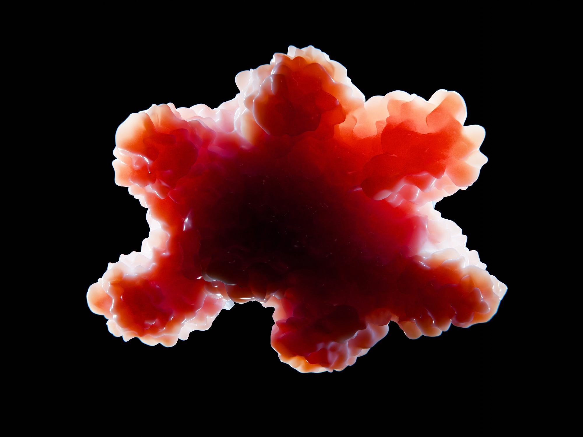
Organoid tissue. Image Credit: Juan Gaertner / Shutterstock. Study: Developmental xenocortication using human-derived organoids in mice
In a recent study published in the journal Nature, researchers created a xenocortical (XCX) mouse model in which human-derived cortical organoids occupied most of the space normally filled by the mouse cortex. The grafts developed diverse human cortical cell types, connected with the host nervous system, generated organized neural activity, and produced measurable responses to hypoxic injury.
Background
Human brain tissue is difficult to study during development, prompting researchers to use stem-cell-derived cortical organoids as experimental models. These three-dimensional tissues can reproduce aspects of cortical neurogenesis and gliogenesis, but organoids grown in laboratory dishes cannot capture how human neural cells interact with a living nervous system or influence behavior.
Transplanting human cortical organoids into rodents offers one way to study these processes in vivo. Existing approaches, however, leave the host cortex intact, limiting the space available for graft growth and creating competition between human and rodent neural circuits.
The researchers therefore sought to create a mouse model with substantially more cortical space available for human-derived tissue while retaining the ability to measure neural activity, connectivity, behavior, and responses to injury.
About the study
Researchers generated immunodeficient apallial mice by conditionally deleting the sister chromatid cohesion factor Esco2 in cells expressing the dorsal and medial pallium marker Emx1 on a severe combined immunodeficiency (SCID) background. This genetic strategy depleted much of the mouse neocortex and hippocampus.
Human induced pluripotent stem (hiPS) cells were differentiated into cortical organoids. Four organoids, aged 30 to 60 days, were transplanted into apallial mice between 5 and 17 days of age.
The team followed graft growth and organization using brain imaging, molecular profiling, neural tracing, spatial transcriptomics, calcium imaging, and electrophysiological recordings. Behavioral testing assessed locomotion, gait, spontaneous behavior, memory-related performance, social preference, and sensory responses.
The researchers also exposed mice to severe hypoxia to determine whether the human grafts produced measurable cellular and behavioral responses to injury.
Study results
The genetic strategy markedly depleted the dorsal and medial pallium. Magnetic resonance imaging (MRI) showed approximately 50% less brain tissue in apallial mice than in controls.
Single-nucleus ribonucleic acid sequencing (snRNA-seq) generated 880,149 profiles and showed a sevenfold depletion of dorsal pallial-derived glutamatergic neuronal classes, with broad losses among neocortical and hippocampal glutamatergic subclasses. Ventral and lateral glutamatergic pallial populations were comparatively preserved.
Human grafts grew extensively after transplantation. Among 29 mice receiving organoids from three hiPS cell lines, graft survival was 86.2%. MRI showed a 4.7-fold increase in graft volume between 2 and 3 months after transplantation. At 3 months, human-derived tissue accounted for 91.9% of the combined cortical tissue volume.
Neural tracing identified organized human-derived pathways and host inputs from regions including the palaeocortex, thalamus, and pallidum. Diffusion-weighted imaging indicated predominantly dorsoventral organization of tissue microstructure, while sparse human-derived projections extended into the cervical spinal cord.
The grafts also contained layer 5 extratelencephalic (L5-ET) glutamatergic neurons, a projection-neuron population rarely generated in cortical organoids grown in the laboratory. Their proportion was more than three times that reported in previous transplantation approaches.
Von Economo neuron (VEN)-like cells were identified in all three sampled XCX mice. The researchers described these cells as VEN-like based on their morphology rather than confirming that they were fully mature von Economo neurons.
Calcium imaging showed large synchronous bursts of neural activity lasting tens of seconds and recurring every few minutes. Individual events began locally and propagated across the graft within about 100 milliseconds. Local field potential recordings confirmed coherent network-wide bursts, while calcium activity was correlated with orofacial movement.
Despite extensive depletion of the native cortex, control, apallial, and XCX mice showed broadly similar locomotion. Motion sequencing indicated that cortical depletion altered the higher-order organization of spontaneous behavior, while transplantation shifted this behavioral pattern without restoring it to the control state.
Both apallial and XCX mice showed changes in paw coordination. In the Y-maze, control and XCX mice performed above chance, whereas apallial mice did not. This finding did not establish that the human graft restored working memory.
Apallial and XCX mice also showed reduced freezing during aversive trace conditioning and contextual testing. Sociability preference did not differ among the groups, although apallial mice investigated less overall, and none of the groups preferred a novel mouse over a familiar one. Mechanical and thermal sensitivity were similar across groups.
The researchers then tested whether xenocortication could provide measurable responses to brain injury. Following a gradual reduction in atmospheric oxygen, mice were exposed to 5% oxygen for 5 hours.
Hypoxia-inducible factor 1 alpha (HIF1α) was detected prominently in the human graft immediately after exposure but was not appreciable in adjacent mouse palaeocortex or similarly exposed control mice. Susceptibility-weighted imaging later detected increased hypointense vasculature in injured XCX mice.
Within the graft, microglial and astroglial density and complexity increased after hypoxia. XCX mice also showed the largest increase in three- to four-paw support during CatWalk gait testing, without corresponding changes in running speed or step cadence.
Conclusions
The study established xenocortication as a platform in which human-derived cortical tissue can occupy most of the cortical space created by genetic depletion in mice. The grafts developed diverse cortical cell types, formed extensive anatomical connections with host tissue, and generated organized spontaneous electrical activity.
The researchers cautioned that anatomical connectivity does not demonstrate pathway-specific functional integration and that it remains unclear whether graft-derived neurons are necessary or sufficient for particular behaviors.
The grafts also remained developmentally immature, with incomplete cortical layering and arealization, limited representation of GABAergic interneurons, immature network properties, and a developmental mismatch between the human graft and rodent host.
The authors emphasized the need for continued ethical oversight as future models become more mature and organized. The present work received approval for animal and stem-cell research and included consultation with bioethicists and an independent ethics committee.
Overall, the platform provides a way to examine developing human cortical tissue within a living nervous system and to link cellular- and circuit-level changes to behavioral readouts.
Journal reference:
- Kaganovsky, K., Kelley, K. W., Gschwind, T., Harary, P. M., Kochalka, J., White, A. D., Lerma-Usabiaga, G., Chen, X., Revah, O., Gore, F., Aoyama, A., Shadrach, J. L., Yoon, S.-J., Valencia, A., Ogawa, S., Reis, N., Vogel, H., Wandell, B., Kaltschmidt, J. A., Soltesz, I., Deisseroth, K., & Pașca, S. P. (2026). Developmental xenocortication using human-derived organoids in mice. Nature. DOI: 10.1038/s41586-026-11032-2, https://www.nature.com/articles/s41586-026-11032-2
 One brain implant could give people with paralysis a more expressive way to communicate