Instead of waiting four weeks to assess wound healing progress, clinicians could one day use a single smartphone thermal image to identify diabetic foot ulcers at risk of non-healing and intervene sooner.
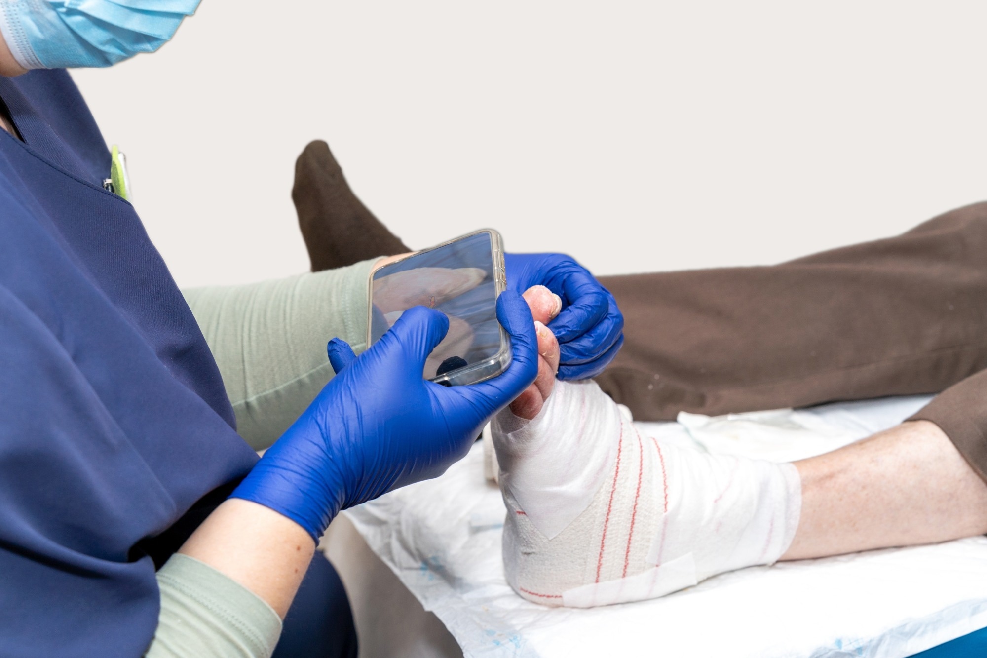 Study: Automated Prediction of Diabetes-Related Foot Ulcer Healing at Initial Assessment. Image credit: Fernando Avendano/Shutterstock.com
Diabetes-related foot ulcers (DFUs) are serious complications of diabetes associated with substantial morbidity and premature mortality. A recent paper in the Journal of Diabetes Science and Technology reports on the preliminary feasibility of an AI-enabled, smartphone-based thermal-imaging platform for use at the first presentation of this condition to predict the potential for healing and thus guide timely management decisions.
Why diabetic foot ulcers need earlier healing predictions
The current clinical marker of a likely chronic diabetes-related foot ulcer (DFU) is a less than 50% reduction in wound area over 4 weeks of standard care. However, this means that a reliable indication of delayed healing based on wound-size reduction becomes available only a month after the first visit, potentially delaying targeted intervention.
Visual inspection and serial wound assessments are typically supplemented by objective measures of limb perfusion and assessments of infection severity and limb threat. Repeated long journeys to specialist foot centers may be taxing for patients in remote areas, in terms of cost, distress, and health status.
Thermal imaging may also be used to identify microcirculatory disturbances and tissue viability, to help improve predictive performance for healing outcomes. However, researchers often compare the temperature of the affected foot with the other foot, which can introduce errors from environmental variables (ambient temperature, humidity, and airflow, for instance) or when both feet are affected, as is common in diabetes.
The authors developed TherMUL, a platform to predict 12-week healing from a single non-contact thermal image taken at the first visit.
AI analyzes thermal patterns within diabetic foot ulcers
TherMUL is a smartphone-based system built around a FLIR One thermal camera. It captures paired thermal and standard color images taken from about 20 cm away. It avoids comparing the affected foot with the other foot.
Instead, it analyzes the texture of the temperature pattern within the wound itself, using gray-level co-occurrence matrix (GLCM) features. During model development, researchers manually identified the wound region on color images, while the preliminary evaluation used automated wound identification. The most important features were energy, homogeneity, and sum of squares (variance). The authors link these to how uniform or uneven the wound’s temperature is.
Uneven temperature may reflect inflammation, infection or poor perfusion. Conversely, homogeneity may reflect uniform tissue metabolism, with less localized ischemia or infection.
Researchers train AI to predict 12-week healing outcomes
The authors conducted a prospective observational cohort study at two specialist foot clinics in Melbourne. Participants were adults with diabetes and peripheral neuropathy with loss of protective sensation. They restricted ulcers to those with low ischemia and infection severity by the Wound, Ischemia and Foot Infection (WIfI) classification.
Baseline images were taken before debridement. The outcome was complete healing at 12 weeks, judged by podiatrists and endocrinologists independently of the model. The predictive model was developed retrospectively using baseline images after the 12-week healing outcomes had been established.
A linear support vector machine was developed using data from 68 ulcers from 56 participants. Of these, 44 healed and 24 did not, including two amputations. The researchers assessed the model’s performance with repeated 5-fold cross-validation. Each of these folds contained about 55 training and 13 test samples.
The training set included approximately 36 healed and 19 non-healed samples. To adjust for the class imbalance, the authors applied the Synthetic Minority Over-sampling Technique (SMOTE) to generate synthetic samples for the unhealed group, enabling oversampling, but only within the training folds.
TherMUL shows moderate accuracy in predicting ulcer healing
During testing, the model had a mean AUC of 0.635, indicating moderate predictive performance. Its sensitivity for detecting unhealed ulcers was 75.8%, and its specificity for identifying healed ulcers was 53%. The mean accuracy was 61%.
The model was therefore better at identifying ulcers unlikely to heal, although it incorrectly classified nearly half of the ulcers that ultimately healed as non-healing. In the training folds, its performance was somewhat higher, with an AUC of 0.717.
A separate set of seven unseen ulcers (four healed and three unhealed) was used to run a preliminary feasibility check. The model correctly classified 5 of 7. The model predicted one healed ulcer would not heal and one unhealed ulcer would heal. However, this small sample precludes definitive conclusions about the model’s performance in other patient populations.
Smartphone imaging could improve access to specialist care
The authors describe the model’s performance as moderate. They say it shows early feasibility of point-of-care prediction at the first visit, without repeated imaging. They present the tool as supporting clinical decision-making and complementing clinician judgment.
According to them, such a tool could be especially useful in rural and remote settings. Notably, GLCM features are interpretable, which may help clinicians understand the predictions and develop appropriate levels of trust in the model.
Moreover, the design offers a potential advantage over earlier thermal imaging tools that rely on longitudinal assessment of changes or asymmetry in temperature at the imaged site over time. This could improve scalability and reduce the burden of repeated imaging on patients.
Small sample and controlled settings limit broader applicability
The model operated under several limitations. The data came only from controlled clinic rooms, which could limit its applicability in home- or community-based settings because variations in ambient temperature, humidity, and airflow were not accounted for.
The authors did not formally assess inter-rater agreement in delineating the wound region. The restriction to neuropathic ulcers with low ischemia and infection severity also limits the generalizability of the findings to more complex DFUs. The sample is small, and the authors call for larger, more diverse datasets with external validation.
Further research is needed to establish whether using TherMUL to guide clinical decisions improves patient outcomes.
AI-powered thermal imaging offers promise for personalized wound care
This study demonstrates the feasibility of predicting DFU healing status using a single image acquired at the initial clinical assessment, representing a substantive advance over existing approaches that rely on longitudinal imaging across multiple follow-up visits.
Future studies should include comparison with established predictors such as WIfI.
In addition, the authors propose that thermal features should be combined with routinely collected variables such as age, diabetes duration, smoking, and neuropathy status to potentially improve predictive accuracy. Finally, they propose that expert-validated multi-level risk grades (namely, low, moderate, and high) for DFU non-healing could increase the clinical relevance of this tool.
 UT MD Anderson announces major expansion to transform cancer care