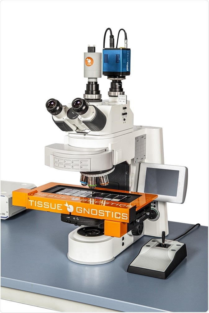
TissueFAXS PLUS is a brightfield system and modular upright fluorescence used for the analysis and scanning of tissue microarrays, smears, cytospins and tissue sections on slides1.

Image credit: TissueGnostics
Its versatility as a whole slide imaging system means it provides a range of capabilities, including multispectral and confocal microscopy, widefield fluorescence and brightfield scanning. The package is completed by inbuilt image analysis software.
Recently, the system has been used for the investigation into what type of immune reactions occur at the site of implanted polypropylene meshes.
Polypropylene meshes
A range of tissue repair procedures use surgical mesh for strengthening and stabilizing soft tissue defects or for supporting prolapsed viscera and organs. Hernia repair is one of their most common applications.
The mesh simultaneously provides a scaffold to support the growth of new tissue while also mechanically strengthening the weakened area; this expands through pores in the mesh until it is eventually surrounded.
Hernia surgery has historically required tissue at the edges of the weakened or torn area to be stitched together. Hernia repair commonly failed because the stitches were frequently subjected to large amounts of strain. There have been considerably fewer hernia recurrences since the introduction of meshes.
Polypropylene was used to make the first meshes; it continues to be the most common type of mesh used in pelvic organ prolapse and hernia repairs.
However, the body raises an immune response to the polypropylene meshes in an attempt to remove them and minimize the potential threat because polypropylene is not a naturally occurring product in the body and is identified as being foreign.
Implant rejection is an ongoing concern with hernia repair despite taking measures to control immune responses to polypropylene mesh implants.
Immune response to implants
When immune cells migrate to the implant site, there is inflammation and a fibrous capsule known as a foreign body granuloma forms. The capsule encapsulates the mesh in order to keep it isolated from the rest of the body2.

Image credit: Yurchanka Siarhei/Shutterstock.com
The activation of the innate immune system is typically considered to be the cause of this granulomatous inflammation in response to a foreign body. It is dominated by macrophages that collect at the mesh surface3,4.
The adaptive immune response involves the release of T-lymphocytes5 and B-lymphocytes and is more specific but takes longer to initiate.
It is possible that adaptive immunity may play a role in the foreign body reaction6,7 because lymphocytes have been detected in the foreign body granuloma that develops at the site of the mesh implant. It is still uncertain what the role of the adaptive immune system at the site of mesh implants is, however.
Evaluation of immune response to polypropylene mesh
The foreign body reaction to seven polypropylene meshes that had been removed from patients due to hernia recurrence8 has been investigated by a recent study. The meshes in the study had been in situ in the abdomen for a median duration of one year.
A TissueGnostics TissueFAXS PLUS system was used to view each mesh by immunofluorescence microscopy to determine which immune cells were present. All specimens showed an apparent pronounced foreign body reaction. The reaction was specific to the area of the mesh fibers where there was dense cellular infiltrates of mononuclear cells.
Regulatory T-cells of the adaptive immune response and T-helper cells were also present, in addition to macrophages from the innate immune system. Additionally, at the mesh fibers, significant clustering of the adaptive immune cells was observed.
The different classes of immune cells’ densities were determined in circles of 1 mm2 across the mesh fibers, in addition to in the surrounding tissue at an increasing distance from the implant’s site.
It was expected that as an immune response against foreign structures 'invading' the body, there would be a high proportion of innate immune cells in the foreign body granuloma, and this proved to be the case.
However, there was a larger number than expected of adaptive immune cells observed. B-cells were found to be clustered around blood vessels serving the tissues surrounding the mesh.
Additionally, the proportions of T cells present were similar to those observed for macrophages. The concentration of both adaptive and innate immune cells showed a significant decrease when the distance from the mesh fibers increased.
This result supports previously stated hypotheses that both adaptive and innate immune cells participate in the chronic foreign body response to implanted polypropylene meshes. Macrophages and T cells are the predominant immune cells involved in this response.
This new finding could reduce the risk of patient morbidity from chronic inflammation at the repair site, rejection and could promote speedy healing by helping physicians control foreign body granuloma formation at the site of hernia repair.
References
- TissueGnostics. Upright Brightfield & Fluorescence Cytometers. https://tissuegnostics.com/products/fluorescence-brightfield-cytometer/tissuefaxs-plus
- Krzyszczyk P, Schloss R, Palmer A, et al. Front Physiol. 2018;9:419.
- Klosterhalfen B, Junge K, Klinge U. Expert Rev Med Devices 2005;2:103–117.
- Pagán AJ, Ramakrishnan L. Annu Rev Immunol. 2018;36:639-665.
- Netea MG, Schlitzer A, Placek K et al. Cell Host Microbe 2019;25:13–26.
- Klinge U, Dietz U, Fet N, Klosterhalfen B. Hernia 2014;18:571–578.
- Tennyson L, Rytel M, Palcsey S et al. Am J Obstet Gynecol 2019;220:187.e1-187.
- Dievernich, A., Achenbach, P., Davies, L. et al. Hernia (2021). https://doi.org/10.1007/s10029-021-02396-7

About TissueGnostics
TissueGnostics (TG) is an Austrian company focusing on integrated solutions for high content and/or high throughput scanning and analysis of biomedical, veterinary, natural sciences, and technical microscopy samples.
TG has been founded by scientists from the Vienna University Hospital (AKH) in 2003. It is now a globally active company with subsidiaries in the EU, the USA, and China, and customers in 30 countries.
TissueGnostics portfolio
TG scanning systems are currently based on versatile automated microscopy systems with or without image analysis capabilities. We strive to provide cutting-edge technology solutions, such as multispectral imaging and context-based image analysis as well as established features like Z-Stacking and Extended Focus. This is combined with a strong emphasis on automation, ease of use of all solutions, and the production of publication-ready data.
The TG systems offer integrated workflows, i.e. scan and analysis, for digital slides or images of tissue sections, Tissue Microarrays (TMA), cell culture monolayers, smears, and other samples on slides and oversized slides, in Microtiter plates, Petri dishes and specialized sample containers. TG also provides dedicated workflows for FISH, CISH, and other dot structures.
TG analysis software apart from being integrated into full systems is fully standalone capable and supports a wide variety of scanner image formats as well as digital images taken with any microscope.
TG also provides routine hematology scanning and analysis systems for peripheral blood, bone marrow, and body fluids.
TG cooperations
TG continuously cooperates with research groups and other companies in the industry to provide novel tools and applications to its customers.
Sponsored Content Policy: News-Medical.net publishes articles and related content that may be derived from sources where we have existing commercial relationships, provided such content adds value to the core editorial ethos of News-Medical.Net which is to educate and inform site visitors interested in medical research, science, medical devices and treatments.