Sponsored Content by BellBrook LabsReviewed by Olivia FrostMay 13 2024
The Transcreener® ADPR Assay is a biochemical HTS test that quantitatively measures the production of ADP-ribose (ADPR) in enzymatic processes. The assay employs a coupling enzyme to transform ADPR into AMP, which is subsequently detected using the far-red competitive fluorescence polarization (FP) assay.
The Transcreener ADPR Assay may identify the activity of human Cluster of Differentiation 38 (CD38) or Poly (ADP-ribose) glycohydrolase (PARG). CD38 regulates cellular NAD homeostasis by degrading NAD and producing ADPR. It has consequences for various pathophysiological diseases, including infection, tumorigenesis, and aging. PARG generates ADPR by the breakdown of poly-ADP-ribose. It is an essential component in DNA damage repair and has been identified as a possible target for anti-cancer therapy.
This article shows how the Transcreener ADPR Assay can offer a robust and dependable tool for discovering CD38 and PARG inhibitors. The selectivity and sensitivity of the assay are demonstrated, followed by its ability to achieve robust assay signals (>100 mP) with less than 10 pM of enzyme and a Z’ value higher than 0.7. The assay was verified in a pilot screen of pharmacologically active compounds.
The Transcreener ADPR FP Assay is an effective technique for discovering CD38 and PARG antagonists, accelerating efforts to regulate these targets pharmacologically.
Transcreener ADPR Assay: ADPR Detection in a Homogenous Format with an FP Readout
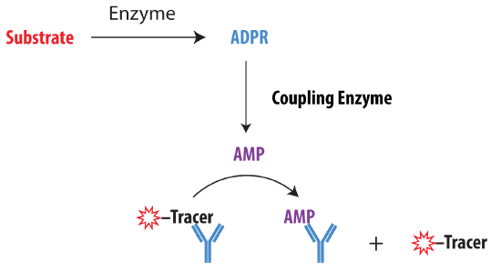
Figure 1. Schematic Overview of the Transcreener ADPR Assay. ADPR produced by the target enzyme is converted to AMP by a coupling enzyme. AMP displaces an AlexaFluor® 633 tracer from the AMP2/GMP2 antibody, resulting in decreased fluorescence polarization. Image Credit: BellBrook Labs
Robust HTS-Ready Assay Procedure
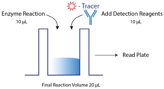
Figure 2. Transcreener ADPR Assay Procedure. The Transcreener ADPR Assay relies on a simple, but robust mix-and-read procedure that is compatible with 96, 384, and 1536-well formats. The target enzyme reaction is run in the presence of coupling enzyme, so that ADPR is converted to AMP in real-time. Detection reagents (AMP2/GMP2 antibody and AlexaFluor 633 tracer) are added along with EDTA to quench the coupling enzyme. Data can then be obtained with a compatible plate reader. Image Credit: BellBrook Labs
Nanomolar Sensitivity and Outstanding Selectivity
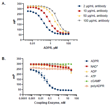
Figure 3. Assay Sensitivity and Specificity of ADPR-AMP Coupling Enzyme. A. Competition curves show detection of ADPR as low as 10 nM and up to 50 μM and illustrate the ability to tune the dynamic range by adjusting AMP2/GMP2 antibody concentration. B. Competition curves show outstanding selectivity of coupling enzyme for ADPR vs. NAD+ , polyADPR, and other nucleotides. Image Credit: BellBrook Labs
Detection of CD38 Under Initial Velocity and Z'
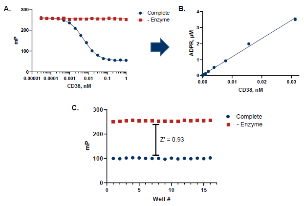
Figure 4. CD38 Assay Robustness for HTS. A. Dependence of assay response on CD38 concentration. B. Conversion of mP to ADPR using a standard curve demonstrates that ADPR formation is linear with enzyme. At 12 pM CD38, about 1.5 μM ADPR was produced, which was 10% substrate conversion from 15 μM NAD+ (approx. Km). C. Z’ measurement using optimized CD38 assay conditions (n=16). A Z’ of 0.93 demonstrates a robust assay method amenable to HTS. Image Credit: BellBrook Labs
Detection of PARG Under Initial Velocity and Z'
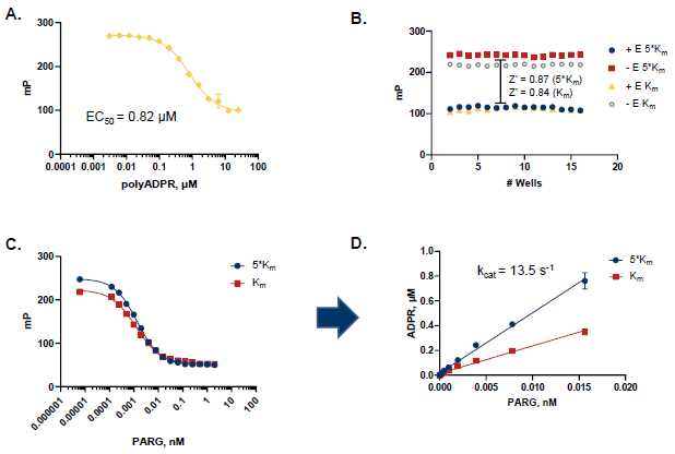
Figure 5. PARG Assay Robustness for HTS. A. polyADPR titration at 7.5 pM PARG; half-maximal response of 0.82 μM (apparent Km). B. Z’ measurement at 5*Km (4.1 μM) or at Km (0.82 μM) concentration of polyADPR and optimal PARG concentration (n=16). A Z’ > 0.8 demonstrates a robust assay method amenable to HTS. C. The competitive curve shows dependence of assay response on PARG concentration at 5*Km or at Km concentration of substrate. D. Conversion of mP to ADPR using a standard curve demonstrates that ADPR formation is linear with enzyme. Image Credit: BellBrook Labs
CD38 Pilot Screen of 1280 Small Molecules
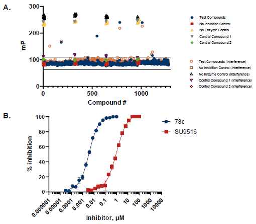
Figure 6. CD38 Pilot Screen and Dose Response. A. 1280 compounds were screened from the Tocris 2.0 Library set. An interference screen was performed to eliminate compounds interfering with coupling enzyme and/or detection reagents. A total of 5 potential inhibitors were identified with polarization values ≥ 3 standard deviations above the mean, in which one showed no interference with assay detection mixture. B. A selected hit from the pilot screen (SU 9516) and the control compound (78c) were tested in dose-response mode with IC50 of 1.01 µM and 4 nM, respectively. Image Credit: BellBrook Labs
PARG Pilot Screen of 960 Small Molecules
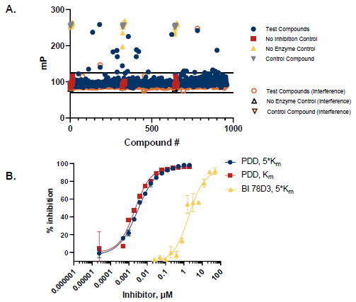
Figure 7. PARG Pilot Screen and Dose Response. A. 960 compounds were screened from the Tocris 2.0 Library set. An interference screen was performed to eliminate compounds interfering with coupling enzyme and/or detection reagents. A total of 15 potential inhibitors were identified with polarization values ≥ 3 standard deviations above the mean, in which 13 showed no interference with assay detection mixture. B. A selected hit from the pilot screen (BI 78D3) and the control compound (PDD 00017273) were tested in dose-response mode with IC50 of 0.84 µM and 1.82 µM at 5*Km concentration of substrate, respectively. IC50 was 1.01 µM for PDD 00017273 at substrate Km, indicating a non-competitive inhibitor. Image Credit: BellBrook Labs
Conclusions
- The Transcreener far-red, FP homogenous immunoassay for ADPR detects product formation by CD38 and PARG at concentrations ranging from 0.01 to 50 µM.
- The assay offers high data quality data (Z’ > 0.7) and signal (>100 mP polarization shift), making it a reliable HTS assay for PARG and CD38.
- Pilot screens validated the assay for discovering CD38 and PARG inhibitors and evaluating IC50 values.
- The Transcreener ADPR Assay can enable quick identification of inhibitors for CD38, PARG, and related ADPR-producing enzymes.
About BellBrook Labs
BellBrook Labs is dedicated to providing scientists with enabling screening tools to accelerate the discovery of more effective therapies. Leveraging its two base platforms, BellBrook has developed easy-to-use assays for hundreds of drug targets.
Transcreener® Biochemical Assay Technology
The proprietary Transcreener HTS Platform uses a highly specific antibody and far-red tracer for fluorescent immunodetection of nucleotides, including ADP, UDP, GDP, AMP, and GMP. Because its based on detection of nucleotides, the assay is universal for use with virtually any enzyme that produces these nucleotides, such as kinases, glycosyltransferases, GTPases, helicases, ATPases, nucleotidases, exonucleases and PDEs. The assay boasts direct detection of many of these enzyme targets (no coupling enzyme needed), simplyifing the protocol and reducing compound interference.
AptaFluor® Biochemical Assay Technology
AptaFluor leverages a spit aptamer technology to directly detect SAH, the common product of Methyltransferases. As the most sensitive HTS methyltransferase activity assay available, AptaFluor dramatically reduces enzyme usage and allows the assay to be run at or below Km for SAM.
Enzolution™ Assay Systems
Enzolution Assay Systems used with Transcreener Assay technology make for a comprehensive assay solution. Enzolution includes the enzyme, substrate, assay plates and buffers required to produce the enzyme reaction. Using these together simplifies researchers' assay needs without the need to spend time and money sourcing enzymes and developing assays.
Sponsored Content Policy: News-Medical.net publishes articles and related content that may be derived from sources where we have existing commercial relationships, provided such content adds value to the core editorial ethos of News-Medical.Net which is to educate and inform site visitors interested in medical research, science, medical devices and treatments.