Sponsored Content by RefeynReviewed by Olivia FrostJul 17 2024
Mass photometry is a powerful and widely regarded analytical technology that is able to accurately measure the mass of single particles in a solution without the need for labels. This technology is used in a diverse array of applications, including biomolecular function research, structural biology, and bioanalytics.
Image Credit: Shutterstock/fusebulb
What is mass photometry?
Mass photometry is an analytical technique used to investigate biomolecules at the single-particle level. It is able to analyze these particles in their native state and without the need for labels.
Mass photometry works by measuring the light scattered by individual particles, using this signal to quantify these particles and measure their mass (Figure 1).
What is mass photometry used for?
Using mass photometry, it is possible to determine the mass distribution and relative concentrations of all species in a particular sample. This technique can be used to characterize nucleic acids, proteins, and a range of other bioparticles (Figure 2).
Mass photometry can be applied to biomolecules on membranes or in solution membranes,1,2 and is ideal for streamlining and accelerating an array of experimental workflows. These workflows include:
- Sample characterization, including direct assessment of sample homogeneity and purity during expression and purification.3
- Protein oligomerization, including the straightforward validation of protein mono or polydispersity as part of formulation or structural biology studies.3,4
- Biomolecular interactions, including the rapid and streamlined quantification of high-affinity interactions among biomolecules.5,6
- Macromolecular complex assembly, including monitoring the assembly and disassembly of complexes over a period of time.7
Mass photometry provides users with quantitative and easy-to-interpret results, meaning it is an ideal solution to complement existing analytical technologies or provide unique insights or confirmation where required.
Key benefits of mass photometry
Mass photometry offers a number of benefits to its users, including:
- The ability to accurately measure the true native behavior of molecules
- Suitability for use in solution and in a range of buffers
- Compatibility with membrane proteins
- A label-free technique with no need for sample modification
- The ability to provide details of all sub-populations in samples
- The capacity for single-molecule counting
- Suitability for working with a wide mass range and high dynamic range
- The ability to use a single assay format while delivering multiple results
- The option to assess homogeneity, structural integrity and activity
- A rapid, straightforward, and user-friendly approach
- A technique that uses a minimal amount of sample
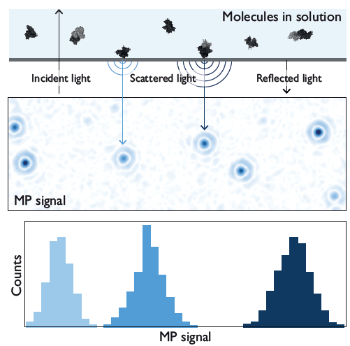
Figure 1. The principle of mass photometry. The light scattered by a molecule that has landed on a measurement interface interferes with light reflected by that surface. The interference signal (MP signal) scales linearly with the molecule’s mass. Image Credit: Refeyn Ltd.
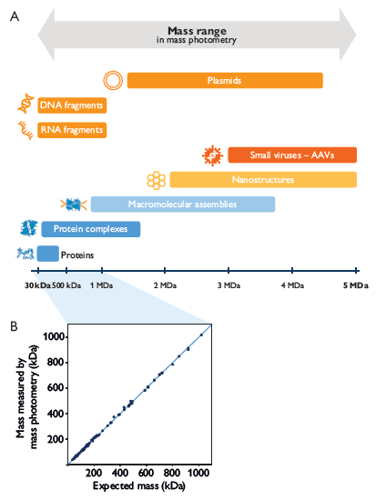
Figure 2. Mass photometry accurately measures diverse biomolecules. A. Mass photometry works across a broad mass range and for different types of molecules. B. The mass measured by mass photometry agrees closely with the expected mass for a range of proteins, illustrating its accuracy. Image Credit: Refeyn Ltd.
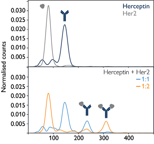
Figure 3. The TwoMP can be used to quantify molecular interactions. The monoclonal antibody Herceptin (trastuzumab) and its target, Her2, were measured individually and in mixtures, demonstrating the instrument’s ability to quantify the interactions of individual antibody molecules with target molecules. mage Credit: Refeyn Ltd.
Mass photometry quantifies binding and assembly
Mass photometry allows users to undertake quantitative studies of biomolecular interactions in solution, such as protein-protein or DNA-protein. As it is able to determine the relative concentrations of both bound and unbound species (Figure 3), mass photometry is ideally suited to the monitoring of interactions and the determination of binding constants for high-affinity interactions.5
Mass photometry’s ability to readily detect sub-species means it is able to analyze complex, multistep interactions with ease. As users are able to determine intermediate binding constants,8 they can optimize experimental conditions as appropriate.
Its broad mass range (Figure 2) also makes it ideally suited to measuring the assembly or disassembly of large, multicomponent complexes (Figure 4) and when looking to follow assembly kinetics.7,8
Mass photometry is useful for quality control
Sample homogeneity is informed by molecular mass, and this universal readout also provides information on the structural integrity and activity of biomolecules and biomolecular complexes.
Using mass photometry, it is possible to ascertain the complete mass distribution of a sample in mere minutes while simultaneously maintaining a near-native aqueous environment for the sample.
Mass photometry is a rapid technique that requires very little sample, making it well suited for applications requiring frequent sample characterization, the highest quality results, and straightforward, streamlined analytical workflows.3
These characteristics make mass photometry an ideal choice for quality control applications such as protein purification optimization and the preparation of complex samples for analysis by cryoEM.3 It is also well suited to the assessment of biomolecular oligomerization behavior, allowing users to rapidly optimize buffer conditions, for example.4
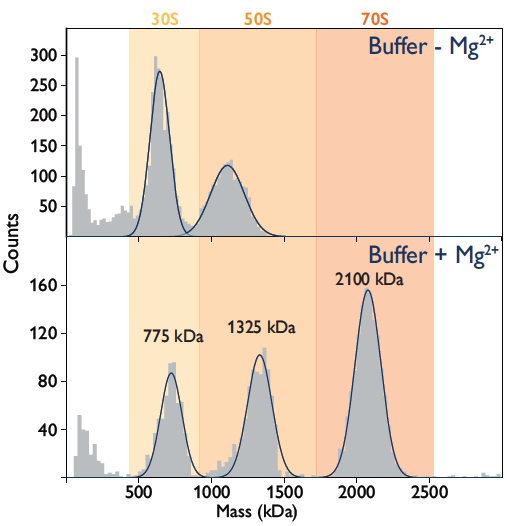
Figure 4. The assembly of bacterial ribosomal complexes monitored. Mass distributions show that ribosome complexes disassemble completely into two subunits in the absence of Mg2+ (top). Complexes assemble in the presence of Mg2+ (bottom). Image Credit: Refeyn Ltd.
How does mass photometry work?
Mass photometry uses light to weigh single molecules. This technique works because the amount of light scattered by molecules correlates with their mass and this scattering of light scales linearly with the particle’s volume and refractive index.
Biomolecules’ optical properties and density generally vary by just a few percent, meaning that the scattering signal will be directly proportional to their mass.
In order to accurately detect the tiny amount of light scattered by individual molecules, mass photometry leverages a combination of carefully controlled illumination, novel spatial filtering in the path of the detection beam,9 and advanced image analysis.8
Mass photometry was developed at Oxford University, building on the underlying principles of interference reflection microscopy10 and interferometric scattering microscopy.11
References and further reading
- Foley et al., Nat Methods 2021
- Heermann et al., Nat Methods 2021
- Sonn-Segev et al., Nat Comm 2020
- Haeussermann et al., Angew Chem Int 2019
- Wu et al., Anal Biochem 2020
- Soltermann et al., Angew Chem Int Ed 2020
- Malay et al., Nature 2019
- Young et al., Science 2018
- Cole et al., ACS Photonics 2017
- 1Verschueren, J Cell Sci 1985
- Ortega-Arroyo et al., Phys Chem Chem Phys 2012
Acknowledgments
Produced from materials originally authored by Refeyn Ltd.
About Refeyn Ltd.
Refeyn are the innovators behind mass photometry, a novel biotechnology that allows users to characterise the composition, structure and dynamics of single molecules in their native environment. We are producing a disruptive generation of analytical instruments that open up new possibilities for research into biomolecular functions.
Spun out of Oxford University in 2018 by an experienced team of scientific professionals, Refeyn aims to transform bioanalytics for scientists, academic researchers, and biopharma companies around the world.
Sponsored Content Policy: News-Medical.net publishes articles and related content that may be derived from sources where we have existing commercial relationships, provided such content adds value to the core editorial ethos of News-Medical.Net which is to educate and inform site visitors interested in medical research, science, medical devices and treatments.