This article is based on a poster originally authored by Jussi Toivanen, Yajuvinder Singh, Leena Rauhala, Taina-Kaisa Stenius, Kimmo Lehtimäki, and Susanne Bäck.
AR DNA Binding Protein 34 kDa (TDP-43) pathology has been associated with familial and sporadic forms of amyotrophic lateral sclerosis (ALS). Almost all ALS cases have cytoplasmic TDP-43 aggregates, and TDP-43 mutations cause this devastating neurodegenerative disease.
The rNLS8 model is a twofold transgenic model in which human TDP-43 expression is suppressed by doxycycline (Dox) and has a faulty nuclear localization signal. The study aimed to compare the disease phenotype to the model's previously reported characterization (Walker et al., Acta Neuropathol 130, 2015).
Methods
Animals:
All animal work followed the European Union Directive 2010/63 and was approved by the national Project Authorization Board. Mice lacking the tetO-hTDP-43-ΔNLS transgene (tTA, 6F+6M) were compared to hemizygous rNLS8 double transgenic mice (rNLS8, 6F+10M; JAX ID: 028412).
The IHC analysis was performed on a separate cohort of rNLS8 (6F+5M) and tTA (4F+3M). From birth until disease induction, animals were fed ad libitum Dox-supplemented food (200 mg/kg Envigo).
Readouts:
- Clinical scores and body weights were measured thrice weekly (Figure 1).
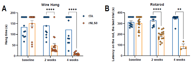
- To test wire hang, invert the top mesh of a wire cage and measure the time it takes to drop (maximum 180 seconds). If the 180-second mark was not met, up to three trials were allowed. The best attempt was chosen for analysis.
- Animals were trained for five minutes at 4 RPM. One hour later, animals were subjected to three consecutive accelerating trials (0 to 40 RPM over 360 s; inter-trial interval 30 minutes).
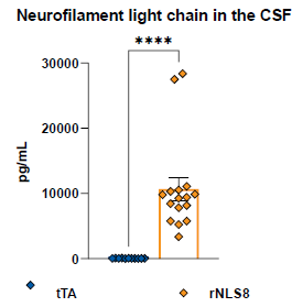
- CSF was analyzed for NfL using the Quanterix SimoaTM NF-Light V2 Advantage test kit (#104073) in single determinations.
- To analyze the neuromuscular junction (NMJ), gastrocnemius muscle cryosections were stained with anti-VAChT and anti-Syp antibodies and AF594-labeled α-bungarotoxin. The number of non-overlapping and overlapping pre- and post-synaptic components was investigated.
- FFPE slices from the lumbar spinal cord were stained with antibodies to identify total TDP-43 (human and mouse) and phosphorylated TDP-43 (Ser409/410). The Visiopharm® system was used for image analysis.

Figure 1. Study design and clinical scoring criteria used in the study. For clinical scoring, animals were lifted by the tail to monitor hind limb clasping and subsequently immobilized to monitor the presence of tremor. Image Credit: Charles River Laboratories.
Results
Clinical scores and body weights: In rNLS8 mice, hind leg weakness began seven days after discontinuing dox (Figure 2A). On day 22, all rNLS8 mice demonstrated complete hind limb clasping (score = 2). After two weeks off dox, rNLS8 mice began to lose weight (Figure 2B). Five of the 10 rNLS8 males reached the study’s endpoints earlier because they lost more than 25 % of their weight. Only one female mouse and no tTA controls met HEP criteria during the study’s follow-up period.

Figure 2. Weakness symptoms and body weight over time. A) Clinical scores as assessed thrice per week, * p < 0.05 (multiple Mann-Whitney U tests). B) Body weights measured thrice per week. Mean±SEM. * p < 0.05 (Mixed-effects ANOVA, Fisher’s LSD). tTA, n=6F+6M; rNLS8, n=6F+10M. The red line indicates the timing of dox diet discontinuation. Image Credit: Charles River Laboratories.

Figure 3. Wire hang times (A) and Rotarod performance (B) at baseline on dox diet and 2 and 4 weeks post off dox. Mean±SEM. **p < 0.01, **** p < 0.0001 (Mixed-effects ANOVA, Fisher’s LSD). Rotarod: tTA, n=6F+6M, rNLS8, n=6F→2F + 10M→1M. Wire hang: tTA, n=6F+6M, rNLS8, n=6F+10M→4. Image Credit: Charles River Laboratories.
NfL: NfL levels in CSF samples collected four weeks after dox removal or at the humane endpoint indicate severe axonal damage and neurodegeneration in rNLS8 animals (Figure 4).

Figure 4. NfL levels in the CSF. Mean±SEM. **** p < 0.0001, Welch’s t-test. tTA, n=5F+6M, rNLS, n=6F+10M. Image Credit: Charles River Laboratories.
NMJ analysis: There was considerable NMJ disintegration in rNLS8 mice, as the fraction of entire NMJs, i.e., with totally or partially overlapping pre- and post-synaptic components, was dramatically reduced when compared to fully intact NMJs in tTA control mice (Figure 5). The percentage of non-overlapping presynaptic and postsynaptic staining was 3.0% in tTA controls and 40.7 % in rNLS8 animals.

Figure 5. A) The proportion of innervated and denervated motor end plates (AChRs) in the gastrocnemius muscle. tTA, n=6F+6M, rNLS, n=6F+10M. Mean±SEM. **** p < 0.0001, Welch’s t-test. B) Example IHC images. All visible NMJs in the tTA example have completely overlapping pre- and postsynaptic components, whereas completely (c), partially (p), and non-overlapping/denervated (n) NMJs are visible in the rNLS8 example. Image Credit: Charles River Laboratories.
TPD-43 pathology: In the spinal cords of rNLS8 animals, the total intensity of TPD-43 staining rose significantly. TDP-43 levels were also much lower in the nucleus than in the cytoplasm (Figures 6A and 6B) showing mislocalisation of this protein. Enhanced phosphorylation of the protein was observed (Figures 6C and 6D).

Figure 6. A) Total TDP-43 IHC staining in the lumbar spinal cord as a ratio of nuclear vs. cytoplasmic intensity. Mean±SEM . tTA, n=3F+2M, rNLS8, n=2F+3M. **** p < 0.0001, Welch’s t-test. B) Total TPD-43 example IHC images from a tTA and rNLS8 mouse. C) pTDP-43 staining intensity in the lumbar spinal cord. ** p < 0.01, Welch’s t-test. D) pTDP-43 example IHC images from a tTA and rNLS8 mouse. Image Credit: Charles River Laboratories.
Summary
All rNLS8 mice demonstrated weakness, weight loss, and poor motor coordination. NfL assays from CSF samples confirmed vast neuronal damage. Neuromuscular endplate denervation was found in the gastrocnemius muscle. TDP-43 pathology was seen in the cytosolic buildup of TDP-43 and enhanced phosphorylation. All symptoms and pathologies investigated in this mouse model are also found in ALS patients, demonstrating the utility of this model for ALS drug discovery.
About Charles River Laboratories
At Charles River, we are passionate about our role in improving the quality of people’s lives. Our mission, our excellent science and our strong sense of purpose guides us in all that we do, and we approach each day with the knowledge that our work helps to improve the health and well-being of many across the globe.
Charles River provides essential products and services to help pharmaceutical and biotechnology companies, government agencies and leading academic institutions around the globe accelerate their research and drug development efforts.
As a fully integrated partner, Charles River can support your research at any point along the drug discovery continuum.
Sponsored Content Policy: News-Medical.net publishes articles and related content that may be derived from sources where we have existing commercial relationships, provided such content adds value to the core editorial ethos of News-Medical.Net which is to educate and inform site visitors interested in medical research, science, medical devices and treatments.