Jun 15 2009
A prototype breast imaging system combining positron emission tomography (PET) and magnetic resonance imaging (MRI) technologies could greatly improve breast cancer imaging capabilities, according to researchers at SNM's 56th Annual Meeting.
Although the system has not yet been tested on humans, initial results from the prototype indicate the system produces a fusion of detailed PET and MRI images that should allow a more accurate classification of lesions in the breast.
"PET and MRI systems are both powerful, noninvasive tools for detecting breast cancer and evaluating treatment, but each of them also has weaknesses," said Bosky Ravindranath, research assistant working with Dr. David Schlyer at Brookhaven National Laboratory, Upton, N.Y., and lead author of a study on preliminary testing of the prototype. "We believe that combining PET and MRI in a single system will eventually yield highly sensitive and specific breast cancer examinations while at the same time compensating for the shortcomings that exist when using only PET or only MRI." 
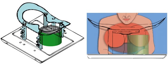
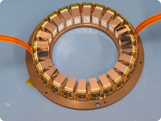
When completed, the dedicated breast PET-MRI system will consist of a modular 3D tomographic PET scanner that is inserted inside a dedicated breast MRI coil produced by Aurora Technologies, Inc allowing both PET and MRI images to be taken simultaneously. The modularity of the PET system would allow for the scanner diameter to be adjusted according to patient breast size. Researchers expect the combined modality scanner will provide anatomical information from the MRI to enhance the resolution provided by PET. At the same time, the predictive power of PET in identifying the type of tumor should be able to overcome MRI technology's traditionally high false-positive rates.
Based on the positive preliminary results, researchers expect to begin testing the system shortly with breast cancer patients. 
Scientific Paper 249: B. Ravindranath, S. Junnarker, S.H. Maramraju; S. Southekal, M. Purschke, S. Stoll, D. Tomasi, P. Vaska, C. Woody, D. Schyler, Department of Biomedical Engineering, Stony Brook University, Stony Brook, N.Y.; and Brookhaven National Laboratory, Upton, N.Y. "Initial results from the BNL dedicated simultaneous PET-MRI breast imaging system prototype," SNM's 56th Annual Meeting, June 13-17, 2009.
http://www.bnl.gov