Vaccines formulated from adenoviral vectors, or mRNAs encoding severe acute respiratory syndrome coronavirus 2 (SARS-CoV-2) spike (S) protein, protect against coronavirus disease 2019 (COVID-19) and permit an effective strategy for combating the COVID-19 pandemic. Vaccines such as these possess the ability to induce the production of neutralizing antibodies within vaccinated individuals. The S protein plays a key role in viral entry into host cells, which is inhibited by these neutralizing antibodies.
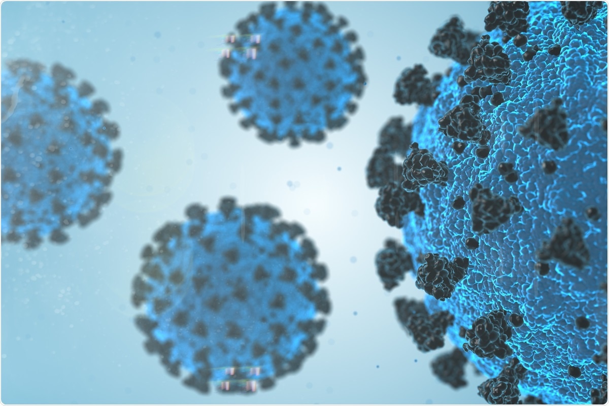
 Study: B.1.617.2 enters and fuses lung cells with increased efficiency and evades antibodies induced by infection and vaccination. Image Credit: Immersion Imagery/ Shutterstock
The vaccines currently used present the S proteins of viruses that were in circulation early during the pandemic as antigens to the immune system. However, variants of concern (VOCs) have emerged within later stages of the pandemic with mutated S proteins that allow for augmented transmissibility and/or immune evasion.
Between April and May 2021, a large surge of COVID-19 cases was identified in India, caused by a new variant (B.1.617). This new variant then branched off into B.1.617.1 (Kappa variant), B.1.617.2 (Delta variant), and B.1.617.3 variants. In a study published in Cell Reports, researchers addressed the question of using reporter particles pseudotyped with the S protein of SARS-CoV-2, which are suitable tools to examine SARS-CoV-2 neutralization antibodies.
The study
Firstly, the authors examined whether the Delta variant S protein mediates entry into cell lines used frequently in research into SARS-CoV-2, including Vero, 293T, Caco-2, and Calu-3 cells, all of which express endogenous angiotensin-converting enzyme 2 (ACE2). The authors observed that the Dela S protein did mediate cell entry into Vero cells and 293T cells with the same efficacy as the original wild-type strain.
The S protein can also drive the fusion of neighbouring cells, which results in multinucleated giant cells (syncytia). This has been seen in vitro, following directed S protein expression or in infected individuals and post-mortem tissues retrieved from patients who died from COVID-19 related complications.
Due to SARS-CoV-2 protein-driven syncytium being linked to COVID-19 pathogenesis, the authors examined the Delta variant S protein's ability to drive cell-to-cell fusion. Interestingly, the authors found that the Delta variant caused larger and more numerous syncytia compared to the wild-type virus.
The authors determined whether recombinant antibodies could inhibit delta variant cell entry. The Delta S protein was observed to be inhibited by three out of four antibodies tested. The only antibody that the Delta variant was not susceptible to was bamlanivimab, which suggests that this antibody is not suitable for treating Delta variant associated SARS-CoV-2 infections.
Finally, the authors tested whether antibodies generated from vaccines or previous infections could inhibit Delta variant cell entry. Plasma from previously infected individuals appeared to inhibit Delta variant S protein with slightly less efficacy when compared to the wild type S protein. Similar results were observed with vaccinations. However, immune evasion was more prominent when compared to convalescent sera.
Implications
This study demonstrates that the Delta variant exhibits immune evasion, enhanced cell entry, and augmented syncytium formation. The findings on the Delta variant's capability of evading antibody-mediated neutralization agree with previous research, although it is more prominent than recently seen.
It is also shown in this study that treating COVID-19 with bamlanivimab alone will not be effective. Still, the data suggest that imdevimab, casirivmab, and etesevimab are effective treatment options, specifically if administered early.
The findings that the Delta variant spike protein is capable of more cell-to-cell fusion when compared to the wild type suggests that more tissue damage may be caused. Thus suggesting the Delta variant is more pathogenic than previous variants and that viral spread through syncytium formation may contribute to the efficient inter-and intra-host spread of this variant.