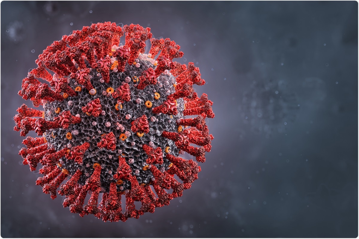
Most early treatments against the coronavirus disease 2019 (COVID-19) have targeted the severe acute respiratory syndrome coronavirus 2 (SARS-CoV-2) spike protein, as it is required for viral entry into host cells. However, many SARS-CoV-2 variants exhibit significant mutations in the spike protein, thus reducing the effectiveness of many vaccines and monoclonal antibody treatments that target this antigen.

Study: Human Galectin-9 Potently Enhances SARS-CoV-2 Replication and Inflammation In Airway Epithelial Cells. Image Credit: Corona Borealis Studio / Shutterstock.com

 This news article was a review of a preliminary scientific report that had not undergone peer-review at the time of publication. Since its initial publication, the scientific report has now been peer reviewed and accepted for publication in a Scientific Journal. Links to the preliminary and peer-reviewed reports are available in the Sources section at the bottom of this article. View Sources
Study findings
In the current study, Calu-3 human airway epithelial cells (AECs) were dosed with recombinant Gal-9 to determine the 50% cytotoxic concentration (CC50) using the MTT assay. Calu-3 cells were then dosed with 50 nanomolar (nM), 100nM, or 250nM Gal-9 for six hours before viral infection with SARS-CoV-2. The cells then remained in the media for 24 hours before SARS-CoV-2 infection was measured by quantitation of nucleocapsid (N) gene expression.
As the Gal-9 concentration increased, SARS-CoV-2 infection increased significantly in a dose-dependent manner, with the highest concentrations showing up to 27-fold increases. The release of infectious virus into the supernatant increased in a similar manner, as measured by the median tissue culture infectious dose (TCID50). Immunofluorescence assays with specific staining of the N protein confirmed the enhancement of virus production by Gal-9.
The researchers then investigated the specific stage of the replication site impacted by Gal-9 by examining Calu-3 cells treated with 350 nM Gal-9 before and after SARS-CoV-2 infection. To this end, pre-treated cells showed significantly higher virus production, thus suggesting that Gal-9 impacts the early stage of the viral life cycle.
![Gal-9 increases virus production in SARS-CoV-2-infected Calu-3 cells. (A) Cellular toxicity was examined in Calu-3 cells using an MTT assay and was expressed as relative cell viability as compared to Gal-9-untreated control (set at 100%). The LogCC50 value for Gal-9 is displayed. The red arrows represent 250 nM and the CC50 value (597 nM) of Gal-9, respectively. (B) The effect of Gal-9 on viral N gene expression in Calu-3 cells was measured by RT-qPCR. Cells were pretreated with Gal-9 at the indicated concentrations for six hours, followed by infection with SARS-CoV-2 (MOI=0.01) for 24 h in the presence of Gal-9. 24 hpi, cells were collected for RNA isolation and RT-qPCR targeting the N gene. (C) Infectious virus release in the supernatant of SARS-CoV-2-infected Calu-3 cells treated with varying doses of Gal-9 as described in (B) were measured using TCID50. (D) Immunofluorescence staining of Calu-3 cells with DAPI (blue) or anti-N Ab (green) pretreated with Gal-9 at the indicated concentrations, followed by infection with SARS-CoV-2 as described in (B). Scale bar, 500 μM. (E) Quantification of SARS-CoV-2 infected cells in Calu-3 cells (shown in panel D). Data are representative of the results of three independent experiments (mean ± SEM). Statistical significance was analyzed by t test. p≤0.05 [*], p≤0.01 [**], p≤0.001 [***], p≤0.0001 [****].](https://www.news-medical.net/image-handler/picture/2022/3/F1.large-108.jpg)
Gal-9 increases virus production in SARS-CoV-2-infected Calu-3 cells. (A) Cellular toxicity was examined in Calu-3 cells using an MTT assay and was expressed as relative cell viability as compared to Gal-9-untreated control (set at 100%). The LogCC50 value for Gal-9 is displayed. The red arrows represent 250 nM and the CC50 value (597 nM) of Gal-9, respectively. (B) The effect of Gal-9 on viral N gene expression in Calu-3 cells was measured by RT-qPCR. Cells were pretreated with Gal-9 at the indicated concentrations for six hours, followed by infection with SARS-CoV-2 (MOI=0.01) for 24 h in the presence of Gal-9. 24 hpi, cells were collected for RNA isolation and RT-qPCR targeting the N gene. (C) Infectious virus release in the supernatant of SARS-CoV-2-infected Calu-3 cells treated with varying doses of Gal-9 as described in (B) were measured using TCID50. (D) Immunofluorescence staining of Calu-3 cells with DAPI (blue) or anti-N Ab (green) pretreated with Gal-9 at the indicated concentrations, followed by infection with SARS-CoV-2 as described in (B). Scale bar, 500 μM. (E) Quantification of SARS-CoV-2 infected cells in Calu-3 cells (shown in panel D). Data are representative of the results of three independent experiments (mean ± SEM). Statistical significance was analyzed by t test. p≤0.05 [*], p≤0.01 [**], p≤0.001 [***], p≤0.0001 [****].
Cells were then incubated at low temperature for two hours, followed by the detection of any attached particles after triple washing of the cells. Gal-9 treatment showed substantial increases in cell attachment in a dose-dependent manner.
To determine if this observation was dependent on the angiotensin-converting enzyme 2 (ACE2) receptor, cells were treated with a competitive anti-ACE2 antibody shown to block SARS-CoV-2 pseudovirus infection. This agent successfully blocked Gal-9 enhanced infection as well.
To determine the mechanisms responsible for the enhancement of infection, cell surface ACE2 and transmembrane serine protease 2 (TMPRSS2) expression were measured by flow cytometry. To this end, Gal-9 did not appear to affect the expression of ACE2 nor TMPRSS2.
Sandwich enzyme-linked immunosorbent assay (ELISA) was then used to examine the binding between ACE2 and the spike protein. This experiment showed significantly enhanced binding when cells were incubated with Gal-9.
![Gal-9 facilitates the cellular attachment and entry of SARS-CoV-2. (A) Attached SARS-CoV-2 virions on the cell surface were detected by RT-qPCR. Calu-3 cells were pretreated with Gal-9 for one hour, then cells were incubated with SARS-CoV-2 (MOI=0.01) in solutions with or without Gal-9 at the indicated concentrations at 4°C for two hours. Cells were washed three times with PBS and harvested for RNA isolation and RT-qPCR measurement of SARS-CoV-2 N gene expression. (B) Relative infectivity of SARS-2-S pseudotyped virus and VSV-G pseudotyped virus in Calu-3 treated with Gal-9 at the indicated concentrations. Calu-3 cells were exposed to Gal-9 for six hours and then infected with SARS-2-S pseudotyped virus or VSV-G pseudotyped virus in solutions containing Gal-9 at the indicated concentrations. Pseudotyped viral entry was analyzed by luciferase activity 24 hpi. Positive serum predetermined to possess anti-SARS-CoV-2 neutralizing activity was used as a negative control. Luciferase signals obtained in the absence of Gal-9 were used for normalization. (C) Relative infectivity of SARS-2-S pseudotyped virus and VSV-G pseudotyped virus in Calu-3 cells treated with anti-ACE2 Ab at the indicated concentrations. Calu-3 cells pre-incubated with anti-ACE2 Ab (R&D Systems, AF933) at the indicated concentrations, or control antibody (anti-goat IgG (R&D Systems, AB-108-C), 50 μg/ml) were co-administered with SARS-2-S pseudotyped and VSV-G pseudotyped virus. At 24 hpi, pseudotyped viral entry was analyzed by luciferase activity. Luciferase signals obtained using the control antibody (anti-goat IgG, 50 μg/ml) were used for normalization. (D) The effect of anti-ACE2 Ab on Gal-9-enhanced cell entry of SARS-2-S was evaluated by measuring luciferase activity. Calu-3 cells were pretreated with Gal-9 for six hours and then pre-incubated with anti-ACE2 Ab (25 μg/ml) or control antibody for one hour, and cells were inoculated with SARS-2-S pseudotyped or VSV-G pseudotyped virus in a solution containing Gal-9 at the indicated concentrations. At 24 hpi, pseudotyped viral entry was analyzed by luciferase activity. Luciferase signals obtained in the absence of both Gal-9 and anti-ACE2 Ab were used for normalization. Data are representative of the results of three independent experiments (mean ± SEM). Statistical significance was analyzed by t test. p>0.05 [ns], p≤0.05 [*], p≤ 0.01 [**], p≤0.001 [***], p≤0.0001 [****].](https://www.news-medical.net/image-handler/picture/2022/3/F2.large-56.jpg)
Gal-9 facilitates the cellular attachment and entry of SARS-CoV-2. (A) Attached SARS-CoV-2 virions on the cell surface were detected by RT-qPCR. Calu-3 cells were pretreated with Gal-9 for one hour, then cells were incubated with SARS-CoV-2 (MOI=0.01) in solutions with or without Gal-9 at the indicated concentrations at 4°C for two hours. Cells were washed three times with PBS and harvested for RNA isolation and RT-qPCR measurement of SARS-CoV-2 N gene expression. (B) Relative infectivity of SARS-2-S pseudotyped virus and VSV-G pseudotyped virus in Calu-3 treated with Gal-9 at the indicated concentrations. Calu-3 cells were exposed to Gal-9 for six hours and then infected with SARS-2-S pseudotyped virus or VSV-G pseudotyped virus in solutions containing Gal-9 at the indicated concentrations. Pseudotyped viral entry was analyzed by luciferase activity 24 hpi. Positive serum predetermined to possess anti-SARS-CoV-2 neutralizing activity was used as a negative control. Luciferase signals obtained in the absence of Gal-9 were used for normalization. (C) Relative infectivity of SARS-2-S pseudotyped virus and VSV-G pseudotyped virus in Calu-3 cells treated with anti-ACE2 Ab at the indicated concentrations. Calu-3 cells pre-incubated with anti-ACE2 Ab (R&D Systems, AF933) at the indicated concentrations, or control antibody (anti-goat IgG (R&D Systems, AB-108-C), 50 μg/ml) were co-administered with SARS-2-S pseudotyped and VSV-G pseudotyped virus. At 24 hpi, pseudotyped viral entry was analyzed by luciferase activity. Luciferase signals obtained using the control antibody (anti-goat IgG, 50 μg/ml) were used for normalization. (D) The effect of anti-ACE2 Ab on Gal-9-enhanced cell entry of SARS-2-S was evaluated by measuring luciferase activity. Calu-3 cells were pretreated with Gal-9 for six hours and then pre-incubated with anti-ACE2 Ab (25 μg/ml) or control antibody for one hour, and cells were inoculated with SARS-2-S pseudotyped or VSV-G pseudotyped virus in a solution containing Gal-9 at the indicated concentrations. At 24 hpi, pseudotyped viral entry was analyzed by luciferase activity. Luciferase signals obtained in the absence of both Gal-9 and anti-ACE2 Ab were used for normalization. Data are representative of the results of three independent experiments (mean ± SEM). Statistical significance was analyzed by t test. p>0.05 [ns], p≤0.05 [*], p≤ 0.01 [**], p≤0.001 [***], p≤0.0001 [****].
When examining the growth curves of the enhanced replication, expression of the N gene increased significantly within one to three hours post-infection (hpi) in Calu-3 cells as compared to untreated controls. Maximum viral yields in treated cells were detected at 36 hpi as compared to 48 hpi in untreated cells.
Microscopy images showed that SARS-CoV-2-mediated cytopathic effects were also much more pronounced in treated cultures. As SARS-CoV-2 is commonly associated with immune dysfunction, the immune response in both treated and untreated cells revealed that Gal-9 induced the expression of interleukin 6 (IL-6), IL-8, and tumor necrosis factor α (TNF-α) to modest levels before SARS-CoV-2 infection.
However, if the cells were infected with SARS-CoV-2, these same immune responses were induced to much higher levels at earlier time frames. Without Gal-9 treatment, IL-6, IL-8, and TNF-α were induced at 24, 26, and 24 hpi, respectively; however, treated cells showed similar levels at 9, 12 and 9 hours.
Ribonucleic acid sequencing (RNA-seq) analysis on Calu-3 cells infected for 24 hours with or without Gal-9 treatment was used to better understand the transcriptional impact of Gal-9. Differentially expressed gene analysis revealed that only one protein-coding gene of RNU12 showed significantly altered expression following SARS-CoV-2 alone. Gal-9 treatment without infection caused significant modulation of 87 genes. Cells infected in the presence of Gal-9 experienced a significant change in their transcriptome, with 1,094 differentially expressed genes.
Ingenuity Pathway Analysis allowed the enriched pathways corresponding to these genes to be analyzed. This revealed that several pro-inflammatory proteins and pathways were activated in the presence of infected cells treated with Gal-9 including IL-6, IL-8, and JAK-STAT signaling pathways.
Finally, AECs cultured at the air/liquid interface (ALI) were pre-treated with Gal-9 and then infected with SARS-CoV-2 for 36 hours. Productive infection could be seen in all cultures from five different donors. Furthermore, real-time polymerase chain reaction (RT-PCR) revealed that IL-6 expression was induced after infection in treated cells.
Conclusions
The researchers of the current study have demonstrated that Gal-9 enhances the ability of SARS-CoV-2 to replicate within cells and that this is effect likely, at least in part, due to the enhanced spike protein-ACE2 binding. The current study has found that Gal-9 treated cells show higher levels of immune response much earlier than non-treated cells and exhibited significantly altered expression of immunologically active genes.

 This news article was a review of a preliminary scientific report that had not undergone peer-review at the time of publication. Since its initial publication, the scientific report has now been peer reviewed and accepted for publication in a Scientific Journal. Links to the preliminary and peer-reviewed reports are available in the Sources section at the bottom of this article. View Sources
Journal references:
- Preliminary scientific report.
Du, L., Bouzidi, M. S., Gala, A., et al. (2022). Human Galectin-9 Potently Enhances SARS-CoV-2 Replication and Inflammation In Airway Epithelial Cells. bioRxiv. doi:10.1101/2022.03.18.484956
- Peer reviewed and published scientific report.
Du, Li, Mohamed S Bouzidi, Akshay Gala, Fred Deiter, Jean-Noël Billaud, Stephen T Yeung, Prerna Dabral, et al. 2023. “Human Galectin-9 Potently Enhances SARS-CoV-2 Replication and Inflammation in Airway Epithelial Cells.” Journal of Molecular Cell Biology, May. https://doi.org/10.1093/jmcb/mjad030. https://academic.oup.com/jmcb/advance-article/doi/10.1093/jmcb/mjad030/7147575.