Several inflammatory and autoimmune conditions can be treated with significantly lower doses of interleukin 1 (IL-2) as compared to those that are used in the treatment of various cancers. This treatment approach involves the stimulation of a subset of CD4+ T-cells with high amounts of the high-affinity trimeric interleukin-2 (IL-2) receptor, which are otherwise known as regulatory T cells (Tregs).
The genetic association between the IL-2 pathway in type 1 diabetes (T1D) has supported the use of low-dose (LD)-IL-2 immunotherapy to rectify the pathogenic IL-2 pathway malfunction in this autoimmune disease. However, several initial trials have demonstrated that while safe, LD-IL-2 immunotherapy had limited therapeutic benefit and inconsistent effects on Treg induction levels in this patient population.

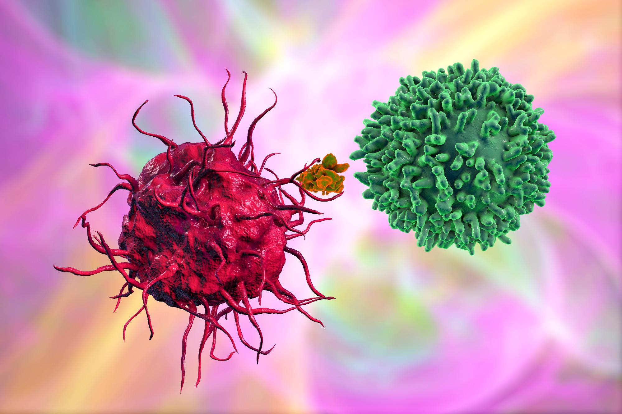
Study: Low-dose IL-2 reduces IL-21+ T cells and induces a long-lived antiinflammatory gene expression signature inversely modulated in COVID-19 patients. Image Credit: Kateryna Kon / Shutterstock

 This news article was a review of a preliminary scientific report that had not undergone peer-review at the time of publication. Since its initial publication, the scientific report has now been peer reviewed and accepted for publication in a Scientific Journal. Links to the preliminary and peer-reviewed reports are available in the Sources section at the bottom of this article. View Sources
Impact of iLD-IL-2 treatment on Teffs and Tregs
In a recent study published on the preprint server medRxiv*, the impact of interval administration of LD-IL-2 (iLD-IL-2) was examined in 18 T1D patients who received a three-day dosing interval with doses ranging from 0.20-0.47x10⁶ IU/m2. Polychromatic flow cytometry (FACS) was then used to characterize cellular changes in blood samples obtained from these patients.
This portion of the study demonstrated that the three-day treatment protocol of iLD-IL2 for a period of one month selectively increases the number of thymic-derived FOXP3+HELIOS+Tregs. Furthermore, this treatment approach did not appear to alter the activity of effector T-cells or CD56dim NK cells.
Of the 18 study participants, blood samples from 12 individuals obtained on Days 0, 24, and 55 were selected for T-reg suppression assays. Herein, 500 CD4+CD25-/loCD127+ effector T-cells (Teffs) which were in the presence of or absence of CD4+CD25highCD127low Tregs at various ratios were sorted and used to determine the percentage of suppression in each culture. Notably, no changes in the in vitro capacity of Teffs were identified following IL-2 treatment as compared to pre-treated cells.
Multiomics findings
A targeted multiomics approach established from the BD Rhapsody™ system was then used in 13 selected samples. This approach allowed the researchers to perform a parallel quantification of specific messenger ribonucleic acid (mRNA) and cell-surface proteins residing within each peripheral blood mononuclear cell (PBMC) isolated from these patients.
A total of 565 mRNA transcripts and 65 surface protein targets were identified at the single-cell level. Fixed proportions of CD4+ Tregs, CD4+ Teffs and CD8+ T-cells, CD56br cells, and NK cells at 30%, 25%, 12%, and 8%, respectively, were isolated from each population. Fifteen distinct functional clusters were identified within the sorted T- and NK-cell populations. Furthermore, a similar representation was observed between the five sorted immune cell populations.
Impact of iLD-IL-2 treatment on FOXP3 and IKZF2 in T-cells
Three distinct clusters of Treg transcription factors were identified that corresponded to different populations of CD127lowCD25hiT-cells. These clusters included CD45RA+FOXP3+HELIOS+, CD45RA-FOXP3+HELIOS+, and CD25+FOXP3-HELIOS- clusters.
Most of the isolated CD127lowCD25hi Treg cells expressed both FOXP3 and HELIOS. Moreover, memory cells expressing both of these transcription factors were associated with a greater level of heterogeneity. Whereas one cluster of CD80+ Tregs expressed a greater level of tissue-homing receptors, another cluster exhibited a higher expression of both human leukocyte antigen (HLA) II and other Treg markers like CD39 and glucocorticoid-induced tumor necrosis factor receptor (TNFR)-related protein (GITR).
Comparably, a subset of CD45RA+ cells was identified in CD127lowCD25hi Treg cells that expressed low levels of both FOXP3 and HELIOS. These CD45RA+ cells likely upregulate CD25, thereby contributing to an increased expression of IL-2 in these cells.
Impact of iLD-IL-2 on NK cells
The researchers also sought to investigate the impact of iLD-IL-2 on a subset of NK cells that expressed high levels of CD56 (CD56br NK cells). To this end, an increased frequency of these cells was observed, which was even stronger than the rise in CD4+ Tregs. Taken together, these observations were consistent with the relatively high affinity of CD56br NK cells as compared to the overall NK population.
Comparably, the frequency of CD56dim NK cells, which make up a majority of CD56+ NK cells in circulation, was not altered.
Shared inflammatory gene expression in COVID-19
A total of forty-one genes that were significantly upregulated or downregulated at Day 55, as compared to Day 0, were identified as Day 55 signature genes in the current study. The researchers then sought to identify the expression of these signature genes in samples obtained from 13 patients who had previously recovered from the coronavirus disease 2019 (COVID-19) and compared their results to ten healthy controls.
Notably, the researchers utilized data from the Oxford COVID-19 Multi-omics Blood Atlas (COMBAT) study, wherein 1,419 genes were identified to contribute to COVID-19. Of these genes, 77 were also present in the transcriptional panel identified in the current study.
The researchers used this information to determine how an inflammatory condition like COVID-19 could also alter the immune transcriptional profile. Almost all of the Day 55 signature genes had negative loading scores against COVID-19 and vice-versa, thus indicating a strong inverse correlation between the expression of these genes induced by IL-2 treatment and COVID-19.
Conclusions
The findings from the current study demonstrate that the administration of three doses of iLD-IL-2 over a one-month period can selectively increase the frequency and number of thymic-derived FOXP3+ HELIOS+ Tregs, while having no effect on the proliferation or stimulation of typical Teffs or CD56dim NK cells. Furthermore, this treatment approach led to a significant increase in the proportion of naive FOXP3+ HELIOS+ Tregs, thereby demonstrating that these effects are due to a higher representation of thymic-derived Tregs rather than the proliferation of peripherally-induced Tregs.
In addition to these impacts on Teffs and NK cells, the iLD-IL-2 treatment approach utilized in the current study did not elicit any cytotoxic effects in T-cell gene expression levels. Thus, the Treg-specific effect of this treatment strategy does not lead to an unwanted pro-inflammatory response in treated patients.
Taken together, the current study provides insights into the mechanisms of iLD-IL-2 in T1D patients while also supporting the daily interval dosing approach utilized here as compared to the conventional daily dosing regimen. Simultaneously, the researchers confirm the safety profile of this treatment approach for a potential long-term strategy in the treatment of T1D, as well as other autoimmune diseases.
Notably, the researchers also identified similar physiological mechanisms to exist between both T1D and COVID-19; however, the extent of the differential expression after IL-2 treatment was smaller in T1D patients as compared to those with COVID-19. Although previous studies have demonstrated increased levels of pro-inflammatory mediators in the blood of COVID-19 patients, the findings from this study indicate that the ability of this severe viral infection to induce stable and potentially long-term changes in this gene expression signature.

 This news article was a review of a preliminary scientific report that had not undergone peer-review at the time of publication. Since its initial publication, the scientific report has now been peer reviewed and accepted for publication in a Scientific Journal. Links to the preliminary and peer-reviewed reports are available in the Sources section at the bottom of this article. View Sources
Journal references:
- Preliminary scientific report.
Zhang, J., Hamey, F., Trzupek, D., et al. (2022). Low-dose IL-2 reduces IL-21+ T cells and induces a long-lived antiinflammatory gene expression signature inversely modulated in COVID-19 patients. medRxiv. doi:10.1101/2022.04.05.22273167. https://www.medrxiv.org/content/10.1101/2022.04.05.22273167v1.full-text.
- Peer reviewed and published scientific report.
Zhang, Jia-Yuan, Fiona Hamey, Dominik Trzupek, Marius Mickunas, Mercede Lee, Leila Godfrey, Jennie H. M. Yang, et al. 2022. “Low-Dose IL-2 Reduces IL-21+ T Cell Frequency and Induces Anti-Inflammatory Gene Expression in Type 1 Diabetes.” Nature Communications 13 (1). https://doi.org/10.1038/s41467-022-34162-3. https://doi.org/10.1038/s41467-022-34162-3.