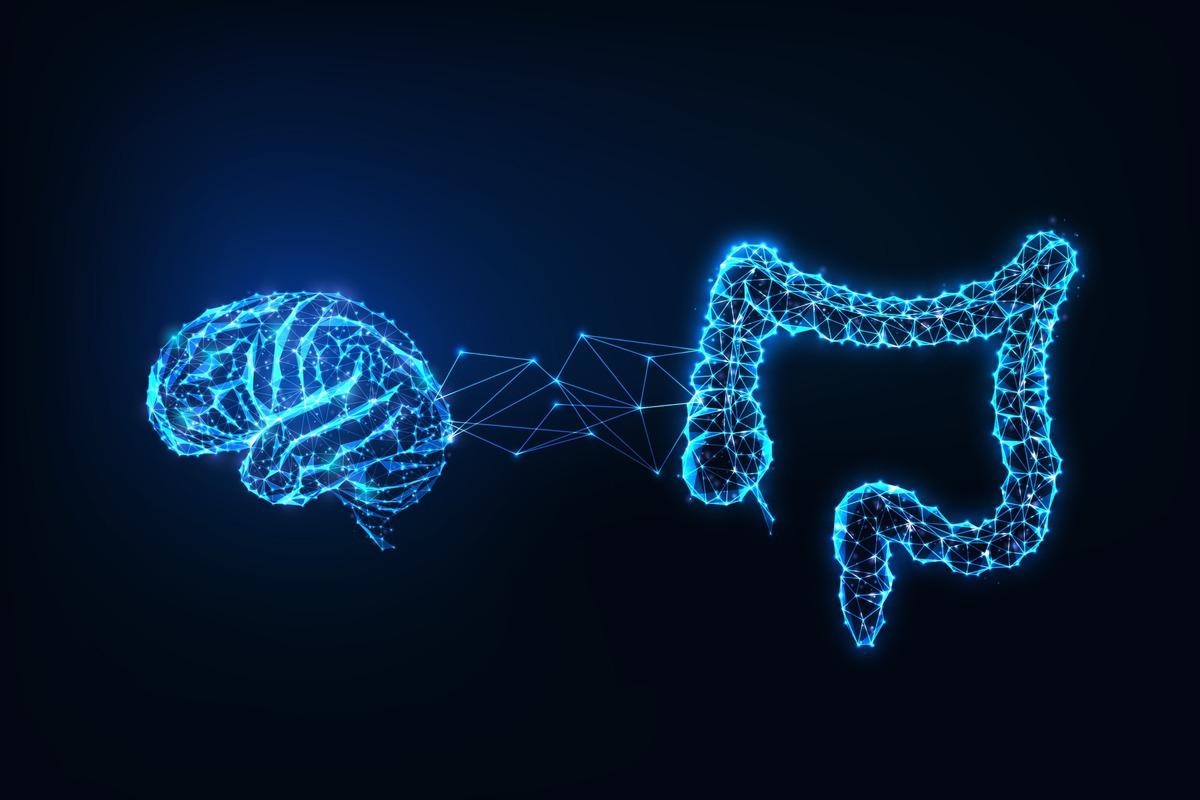
The brain is the central information center and constantly monitors the state of every organ present in a body. Previous research has shown that the brain also receives signals from the gut microbiota.

Study: When the brain feels the bugs. Image Credit: Inkoly / Shutterstock.com
The brain and the gut microbiome
Previous research indicates that structural components from intestinal bacteria can elicit pro-inflammatory responses in the body and, as a result, have an indirect effect on the brain. This phenomenon occurs through peripheral neurons or molecules that are released by immune cells after exposure to bacterial cells circulating in the blood.
In the 2022 Science study, Gabanyi and colleagues discuss microbiome-brain communication. Herein, the researchers report that some neurons in the brain can directly identify bacterial cell wall components and subsequently initiate altered feeding behavior and temperature regulation.
The hypothalamus is a region in the brain that connects the central nervous system (CNS) to the endocrine system through the pituitary gland. Moreover, the hypothalamus regulates various functions such as thirst, hunger, reproduction, sleep, body temperature, and circadian rhythms by inhibiting or stimulating neurons. To date, there is a limited amount of research on how the hypothalamus recognizes the state of the gastrointestinal lumen and perceives the microbes it is harboring.
Commensal microorganisms are typically recognized through pattern recognition receptors (PRRs) of the innate immune system. For instance, NOD2 is involved with the identification of muramyl dipeptide (MDP), which is a peptidoglycan fragment of the bacterial cell wall.
Previous studies have highlighted the functions of NOD2 beyond those which are related to innate immunity. However, the mechanisms responsible for the connection between bacterial peptidoglycans and neuronal functions of the brain remain largely unknown.
What happens when microbial components reach the brain?
Gabanyi and her team addressed this gap in research by studying the NOD2-GFP reporter gene in mice, which helped them investigate the function of NOD2 in different parts of the CNS. Although microglia and endothelial cells were found to express NOD2 in all areas of the brain, NOD2 expression in neurons occurred only in specific regions, such as the striatum, thalamus, and hypothalamus.
The researchers also observed that muropeptides were able to cross the intestinal barrier and reach the systemic circulation system in mice. These peptides were later detected in the brain tissues of all mice. Notably, the extent of their expression was greater in female mice as compared to males.
The researchers also generated a novel mouse model that lacked NOD2 in inhibitory GABAergic neurons (VgatDNod2 mice) and excitatory neurons expressing calcium/calmodulin-dependent protein kinase II (CamKIIDNod2 mice). Aged female VgatDNod2 mice gained weight, had altered body temperature, and increased feeding. These phenotypic events were caused by MDP, as mice treated with MDP exhibited a reduction in food intake as compared to mice that received MDP isomer treatment, which cannot activate NOD2.
The scientists also identified the regions of the brain that were affected by MDP. In this context, they mapped the expression of the neuronal activity marker Fos across different areas of the brain in both male and female mice of varied age groups and treated them with MDP or the control isomer. The arcuate nucleus of the hypothalamus exhibited reduced Fos expression in older female mice compared to males.
Studies have shown that within the arcuate nucleus, the GABAergic population is responsible for food intake, which is constituted by AgRP+ NPY+ neurons. These genes are active during fasting and are silenced upon exposure to food.
Interestingly, Gabanyi et al. observed that these neurons express NOD2 and that MDP exposure suppresses their activity. A decreased activity of GABAergic arcuate nucleus neurons was also identified in both mice.
How does NOD2 expression regulate food intake?
The researchers also infected NOD2fl/fl mice with a Cre-expressing virus in their hypothalamus to locally target NOD2+ GABAergic neurons. Altered phenotypes, such as differential food intake and weight gain in both groups of mice, which included one group treated with MDP and the other with control, returned to normal once treated with broad-spectrum antibiotics.
This finding implies that a decrease in the gut microbiome occurred after antibiotics treatment. This resulted in a reduction in the number of circulating muropeptides that subsequently altered neuronal sensing through its activity on NOD2.
Conclusions
In this study, Gabanyi and her research team highlight the possibility that bacterial components could directly regulate the appetite of individuals. These findings have presented the potential of PRR biology in the brain, which could be exploited to fight against the rising global problem of obesity.