An all-in-one two-photon and/or confocal microscopy system called intravenous microscopy (IVM) was created and refined for in vivo longitudinal imaging of live animal models.
This cutting-edge equipment was created to help biologists and translational scientists understand the underlying mechanisms of all biological phenomena at the tissue and cellular levels. It was designed for ease of use and increased throughput.
Confocal IVM systems allow optical sectioning of in vivo tissue by rejecting out-of-focus fluorescent light from the background tissue, producing high-contrast and high-quality images.
Longer-wavelength near-infrared (NIR) fs-pulse excitation lasers used in two-photon IVM systems can provide label-free, non-linear multi-harmonic generation imaging (SHG, THG) and deep tissue imaging.
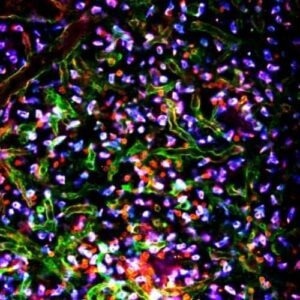
Image Credit: Scintica Instrumentation Inc.
Anesthesia inlet/outlet and body temperature control are fully integrated into the stage of IVM upright systems.
In addition, the system has ultrafast imaging rates to track rapidly moving imaging targets within the tissue and state-of-the-art live tissue motion compensation for better image quality. The system can be set up for two-photon or confocal microscopy modules.
Features and benefits
An all-in-one two-photon and/or confocal microscopy system called intravenous microscopy (IVM) was created and refined for in vivo longitudinal imaging of live animal models.
Integrated heated animal stage and physiological controller
Guarantees the health of the animals during the imaging session and the uniformity of the animals in research.
User-friendly ergonomics and user interface
It makes it simple and enables professionals and non-experts to reproduce results.
Fast to ultrafast scanning
It enables users to monitor many cells' movements in vivo to better understand the biological processes under investigation.
Motion compensation function
Imaging dynamic organs improves image quality by automatically adjusting for the impact of brain pulses and breathing motions.
Integration inhalation anesthesia inlet/outlet
Permits attachment to an external anesthetic inhalation device. All universal anesthetic machines can use it.
4-Color simultaneous imaging
Multiplexity and concurrent observation of multiple labeled tissue components
Animal stabilizing holders and hardware
The animal is quickly and securely stabilized on stage for time-laps and longitudinal imaging to reduce motion artifacts.
4D Imaging
The program allows you to capture 3D stacks of moving objects over time and convert them into 4D images.
Models and specifications
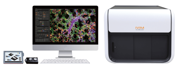
IVM-MS3 Compact Two-Photon Laser Unit. Image Credit: Scintica Instrumentation Inc.
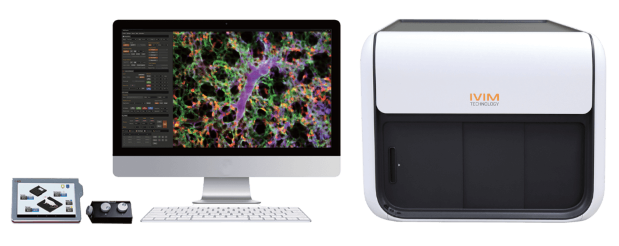
IVM-CMS3 Confocal Laser Unit Compact Two-Photon Laser Unit. Image Credit: Scintica Instrumentation Inc.
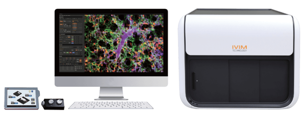
IVM-C3 Confocal Laser Unit. Image Credit: Scintica Instrumentation Inc.
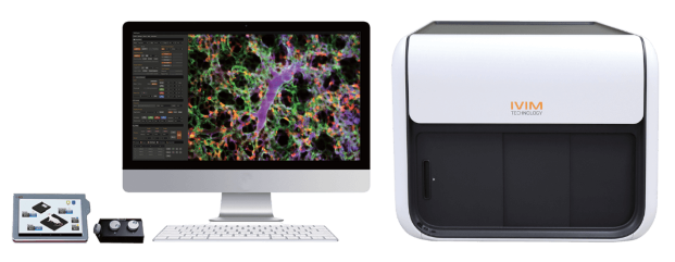
IVM-M3 Tunable Two-Photon Laser Unit. Image Credit: Scintica Instrumentation Inc.
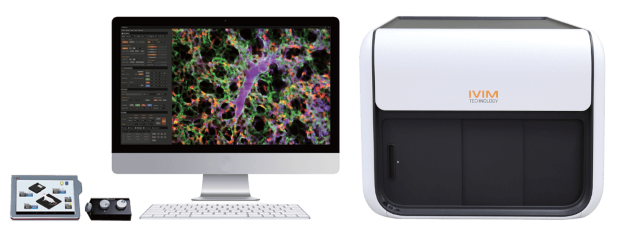
IVM-CM3 Confocal Laser Unit Tunable Two-Photon Laser Unit. Image Credit: Scintica Instrumentation Inc.
Chamber kits
Kits for long-term longitudinal imaging of different organs using in vivo imaging window chambers. Stabilize and expose organs optically for intravital imaging. Enables ongoing imaging for a few weeks or months after surgery.
It encourages moral research methods and reduces the necessity for animal sacrifice after each session. Micro-suction stabilizes tissue movement in the dynamic organ window chamber kit for the uterus, heart, and lungs.
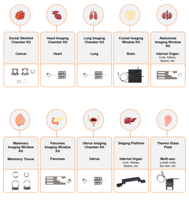
Image Credit: Scintica Instrumentation Inc.
Tissue Motion Stabilizing System (TMS)
- The staging platform offers a practical way to observe several organs in vivo, such as the liver, gut, spleen, and pancreas.
- Imaging the exposed tissue is made flexible and stable by the magnetic base and height-adjustable height.
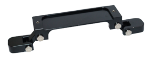
Image Credit: Scintica Instrumentation Inc.
- An organ window chamber that is dynamic Micro-suction is utilized in the lung, heart, and uterine kit to stabilize tissue movement.
- A Tissue Motion Stabilizer (TMS) helps maintain negative pressure between the tissue and the coverslip to prevent tissue from moving during imaging.
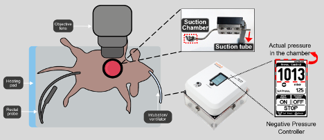
Image Credit: Scintica Instrumentation Inc.
IVIM Tech imaging gallery
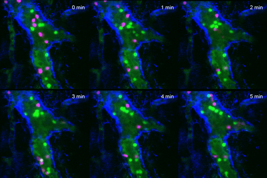
Lymph Node. Image Credit: Scintica Instrumentation Inc.
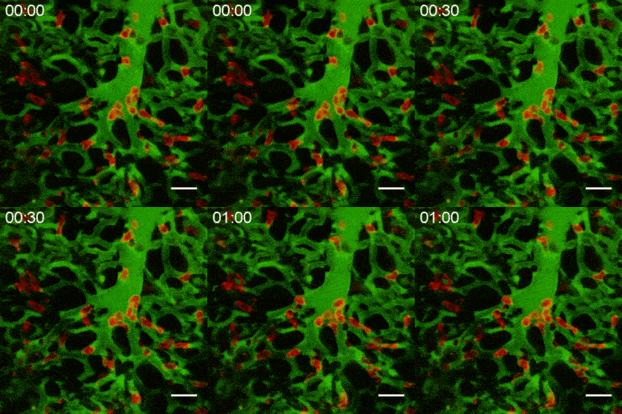
Lung. Image Credit: Scintica Instrumentation Inc.
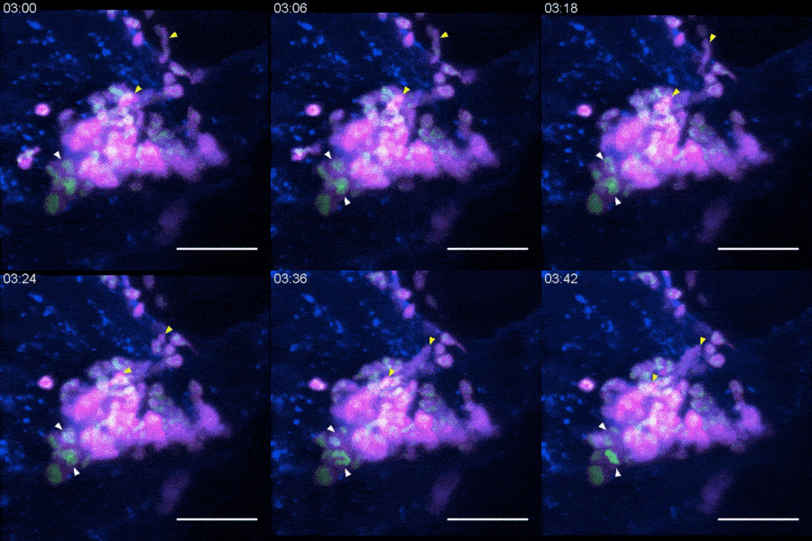
Bone Marrow. Image Credit: Scintica Instrumentation Inc.
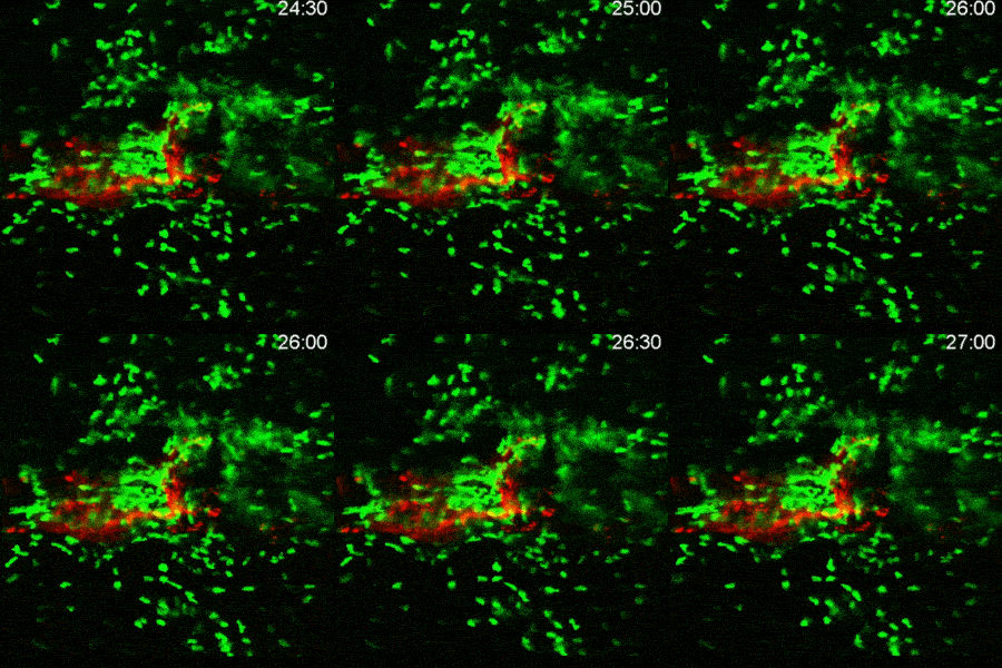
Skin. Image Credit: Scintica Instrumentation Inc.
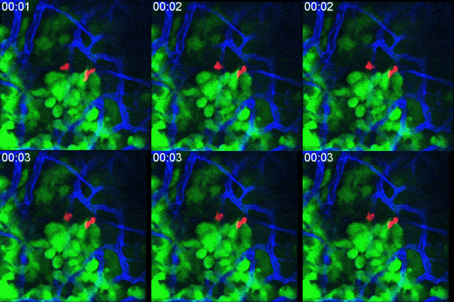
Cancer Xenograft. Image Credit: Scintica Instrumentation Inc.
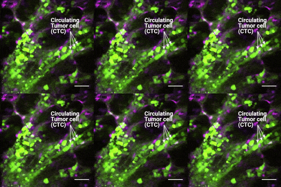
Cancer Metastasis. Image Credit: Scintica Instrumentation Inc.
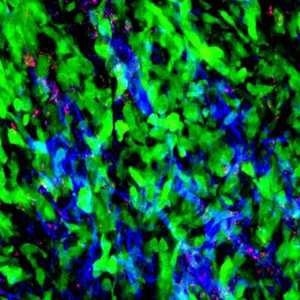
Drug Distribution. Image Credit: Scintica Instrumentation Inc.
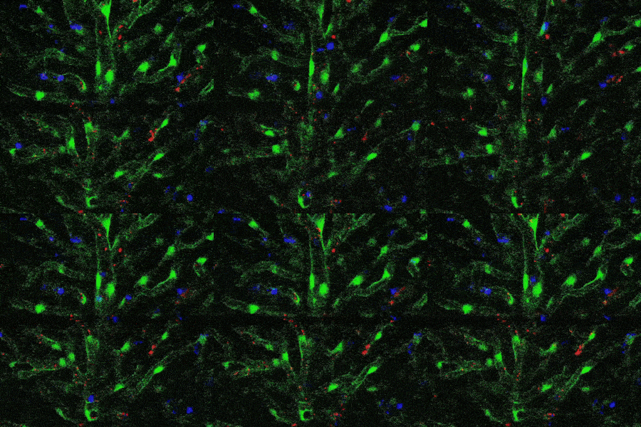
Cancer Drug Delivery. Image Credit: Scintica Instrumentation Inc.
IVIM Series CMS3 2023
IVIM Series CMS3 2023. Video Credit: Scintica Instrumentation Inc.