The Indus Doppler Flow Velocity System is a high-frequency, real-time pulsed Doppler measurement tool with built-in data processing software intended to assess small animals' cardiovascular health.
This system is perfect for examining small animals' quick heart rates and rapid blood accelerations because its high sample rates provide excellent temporal resolution. The Doppler Workstation (DW), Doppler Signal Digitizer (DSD), Pulsed Doppler Transceiver (PDT), a switchable dual channel system operating at 10 and 20 MHz, and a handheld tiny probe or probes are examples of hardware components.
The DSD and the workstation software digitize the PDT's pulsed Doppler signals at high sampling rates. A quick Fourier transform technique processes the obtained signals, and the resulting grayscale Doppler flow velocity spectrograms are shown in real-time.
These spectrograms can be recorded and analyzed using the workstation software, which makes them perfect for creating reports and publishing. Publications have successfully employed this approach using mice, rats, bats, naked mole rats, and other tiny animals. Implanted extra-vascular Doppler cuff probes can also measure blood flow velocities in larger animals.
Non-invasive
A miniature handheld probe can reliably measure flow velocities and differentials in various arteries, including the aorta, by positioning its tip at a sharp angle to the direction of the measured flow.
Small footprint
A strong system that is portable and small enough to be readily scaled in larger facilities or shared between laboratories that collaborate without any issues
Translational data
Numerous research fields, including heart function, myocardial perfusion, pressure overload, arterial stiffness, and more, provide evidence that rodent flow velocity data can be translated into clinical conclusions.
Applications
Cardiac function: Systolic and diastolic
Area
- Heart failure
- Hypertrophy
- Myocardial infarction
- Cardiomyopathy
Flow parameter
- Aortic Exhaustion Rate
- Inflow Velocity of Mitral
Coronary flow reserve
Area
- Pressure Overload-Hypertrophy
- Atherosclerosis
- Myocardial Ischemia
Flow parameter
The ratio of hyperemic to baseline coronary flow velocity
Arterial stiffness (pulse wave velocity)
Area
- High blood pressure
- Atherosclerosis
Flow parameter
- Aortic Arch Speed
- Abdominal Aortic Velocity (Stenosis) Area
Pressure-overload (Stenosis)
Area
TAC Banding Model
Flow parameter
- Carotid peak velocity ratio (R/L)
- Estimating the pressure gradient across stenosis using stenotic jet velocity
Peripheral artery disease and perfusion
Area
Iliac, femoral, carotid, renal, and saphenous veins
Flow parameter
Peripheral vascular flow rates before and following surgery or during a therapeutic response
Transducers
Hand-held transducer
A noninvasive stiff probe with an end-mounted transducer is necessary for most applications. Using epoxy molded into a lens to focus the sound beam, the active element is a 1.0 mm diameter (10 or 20 MHz) piezoelectric crystal recess-mounted at the probe's end.
This targeted probe has been used to noninvasively monitor arterial blood velocity and cardiac blood velocity in mice and rats. The probe can be installed in a micromanipulator, which frequently helps with measurement consistency and precision, or it can be held steadily in the hand.
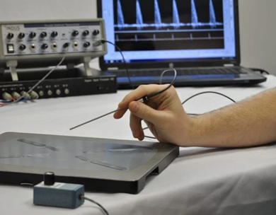
Image Credit: Scintica Instrumentation Inc.
Transducer
A cuff transducer can be employed for extravascular, chronic (implanted) applications, or when a probe cannot reach the vessel. Flexible silicone shapes the cuff body, which is then divided longitudinally to allow it to slide around the vessel. Medical-grade epoxy mounts the 1.0 mm diameter piezoelectric crystal (10 or 20 MHz) at a 45-degree angle.
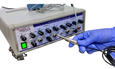
Image Credit: Scintica Instrumentation Inc.
Parameters
Surgical monitoring and vital sign measurements
Peripheral artery: Carotid, renal, femoral, and tail
- Mean and Minimum Flow Velocity
- Index of Pulsatility
- Index of Resistivity
- Peak Velocity
Diastolic: Mitral inflow velocity
- E-stroke velocity and E-peak
- E-time duration
- Time for E-acceleration and E-deceleration
- E-linear deceleration time & rate E-peak to ½ E-peak time
- A-stroke distance
- A-duration of time
- E-A ratio of peak velocity
- Time of isovolumic contraction
- Time spent relaxing isovolumically
Other: Coronary, transverse, and abdominal aorta
- Peak Diastolic Velocity (Coronary)
- Peak Systolic Velocity (Coronary)
- Systolic and Diastolic (Coronary)
- Area Ratios PSV/PDV
- SA/DA Pulse Wave Velocity
Hardware
Doppler Signal Digitizer
- Channels - Channels 1 and 2 = Doppler InPhase & Quadrature Channel 3 = ECG Channels 4 – 8 = Auxiliary inputs
- Range of Input: ±10 V
- Coupling: Choose between AC and DC software
- Sampling: 16-bit sampling at 125 kHz per channel
- Low Pass Filter: 10, 20, 30, 40, 50, 60, 70, 80, 90, 100, 110, 120, 130, 140, or 150 kHz (via hardware)
- Low Pass Filter: 10, 20, 30, 40, 50, 60, 70, 80, 90, 100, 110, 120, 130, 140, or 150 kHz (via hardware)
- High Pass Filter (second or fourth order, via software): 100, 200, 400, 600, 800, 1000, 1500, or 2000 Hz
- Data Link to PC: USB 2.0 (480 Mb/s)
- Digital Signal Processor: 500 MHz Dual Core Processor
- Power: Universal Adapter, 100-240 VAC
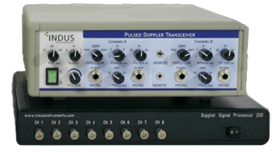
Image Credit: Scintica Instrumentation Inc.
Pulsed Doppler Transceiver
- Channels: Two channels that can be switched between 10 MHz and 20 MHz
- Power: 110 VAC/60 Hz or 220 VAC/50 Hz power
- Audio Outputs - 2 from each channel (InPhase and Quadrature)
- Recorder Outputs - 2 from each channel (Phasic and Mean)
- External Ground - Intended for chasis grounding, if required
- Audio Monitor - Amplifier and speaker selectable from any channel
- Transmitter Pulse Width - 0.4 μs
- USB, RF/DEMOD - Future use
- Variable Range Gate - 1-10 mm (1-13 μs)
- Receiver Pulse Width - 0.32 μs
- Velocity Outputs - 0.25 V/kHz simultaneous Phasic & Mean
- Probe Connection - Floating & differential (single-ended, differential)
- Phasic Output Filter - Phasic (1 pole at 50 Hz), Damped (1 pole at 15 Hz), and Mean (2 poles at 0.25 Hz)
- Velocity Range - 1-100 cm/s at 0° angle, 2-200 cm/s at 60° angle
- Electrical Zero - Front panel switches
- Ultrasound Frequency - 10 MHz // 20 MHz
- Transmitter Output - 25 Vpp into 50 Ohm // 35 Vpp into 50 Ohm
- Controls - Range adjustment, Polarity Switch, Filter
- Pulse Repetition Frequency - 31.25, 62.5, 125 KHz // 62.5, 125 KHz
- Audio Bandwidth - ≈ 100 Hz to 15 KHz // ≈ 200 Hz to 25 KHz
Imaging gallery - Doppler Flow Velocity System
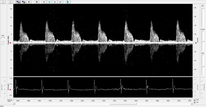
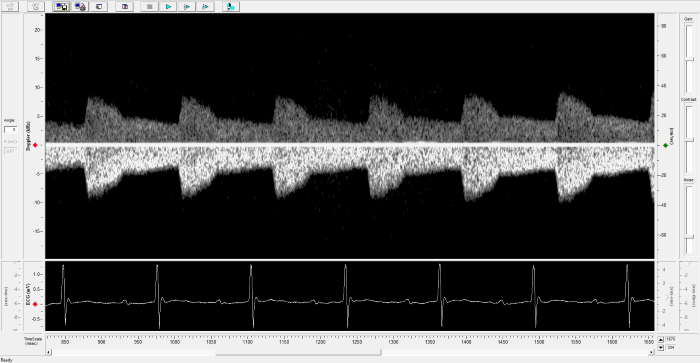
Carotid Image. Image Credit: Scintica Instrumentation Inc.
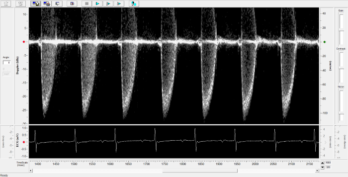
Renal Flow. Image Credit: Scintica Instrumentation Inc.
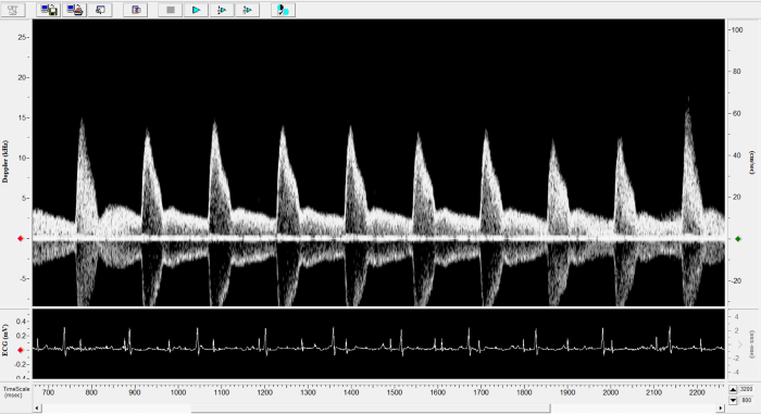
Pulmonary Flow. Image Credit: Scintica Instrumentation Inc.
Mouse - Abdominal Aorta. Image Credit: Scintica Instrumentation Inc.
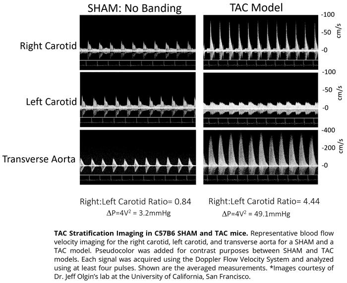
Mouse - TAC Procedure Imaging. Image Credit: Scintica Instrumentation Inc.
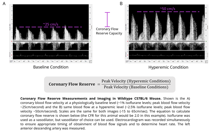
Mouse - Coronary Flow Reserve Imaging. Image Credit: Scintica Instrumentation Inc.
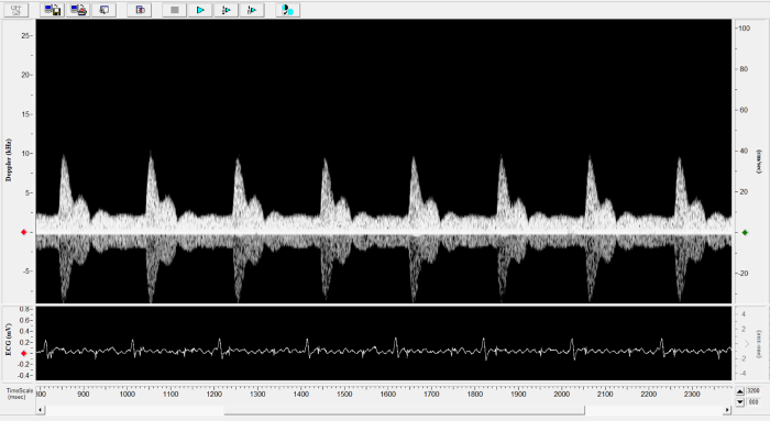
Rat - Left Carotid. Image Credit: Scintica Instrumentation Inc.
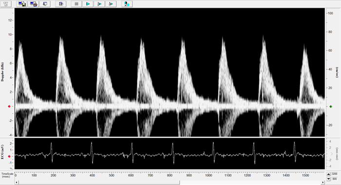
Rat - Transverse Aorta. Image Credit: Scintica Instrumentation Inc.
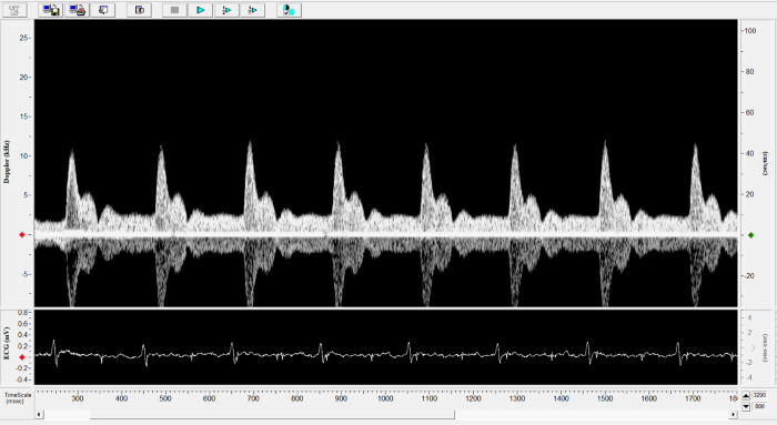
Rat - Right Carotid. Image Credit: Scintica Instrumentation Inc.
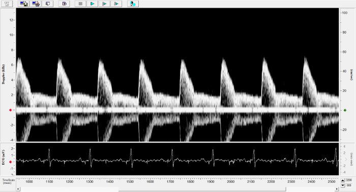
Rat - Abdominal Aorta. Image Credit: Scintica Instrumentation Inc.
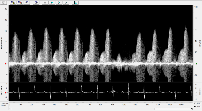
Mouse - Left Anterior Descending Coronary Artery. Image Credit: Scintica Instrumentation Inc.
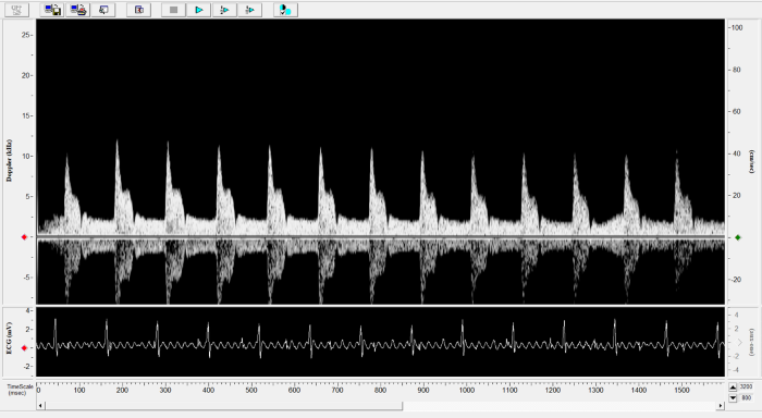
Mouse - Left Carotid. Image Credit: Scintica Instrumentation Inc.
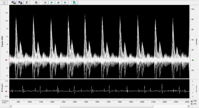
Mouse - Mitral Inflow. Image Credit: Scintica Instrumentation Inc.
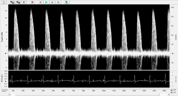
Mouse - Ascending Aorta. Image Credit: Scintica Instrumentation Inc.