The Newton 7.0 is a state-of-the-art optical imaging system that utilizes bioluminescence, fluorescence, and 3D tomographic imaging in one device. Thanks to its sophisticated features and easy-to-use interface, it is perfect for in vitro, ex vivo, and in vivo imaging applications and for imaging numerous specimens at once.
The system has a cutting-edge 4.6-megapixel CCD camera with one of the biggest apertures. This camera's exceptional sensitivity for a range of luciferase enzymes and fluorophores frequently utilized in preclinical research makes fast and effective signal acquisition possible.
Time is saved in longitudinal studies thanks to the user-friendly software and straightforward workflow, which are geared for multi-user use.
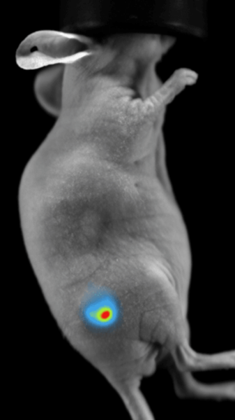
Image Credit: Scintica Instrumentation Inc
Features and benefits
With the user in mind, the Newton 7.0 is a cutting-edge optical bioluminescence, fluorescence, and 3D tomographic imaging system.
State-of-the-art camera technology
- -90 ℃
- 16-bit, scientific grade
- 4.6 MP, native resolution
- CCD Absolute Cooling
- Lens Aperture f/0.7
- 4.8 Optical Density
- 10 MP Image Resolution
Powerful fluorescent excitation
Eight excitation channels in the visible and near-infrared range are included in the Newton 7.0. The illuminating light is closely controlled by two strong Laser Class II arrays, which provide a high-intensity, direct light.
Motorized darkroom with adjustable field-of-view
Thanks to Vilber's clever darkroom architecture, the camera (Z-axis) and animal pad (X/Y axis) may move fully motorized around the macro imaging field of view (6 × 6 cm) and the full field of view (20 × 20 cm) to photograph up to five mice.
Full spectrum tunability
Equipped with 8 excitation channels and 8 emission filters, this system spans the entire spectrum from blue to infrared,
Hard-coated narrow bandpass filters are employed to collect emission wavelengths and limit cross-talk across signals, enabling seamless imaging of all of the most regularly used fluorophores.
3D optical tomography
An integrated 3D tomography module reconstructs bioluminescent signals in 3D and overlays them onto a topographical model of the imaging subject.
To better understand anatomical and deeper tissue features, the mouse organs and bones can be superimposed onto the topographical model using the digital organ and bone library.
License-free acquisition and analysis software
Free upgrades and limitless licenses are included with the easy-to-use program.
Both novice and expert users can swiftly image their subjects by creating custom acquisition procedures or using the factory default acquisition protocols.
Data that complies fully with GLP and CFR21 can be exported in 8-bit.jpg or 16-bit.tiff formats.
Save ROI and analysis templates for high-throughput side-by-side analysis of up to ten images in the analysis module.
Imaging modes
Fluorescence imaging
Vilber's dynamic range of emission filters can be employed in fluorescence imaging to identify fluorescent reporter genes or dyes in vivo.
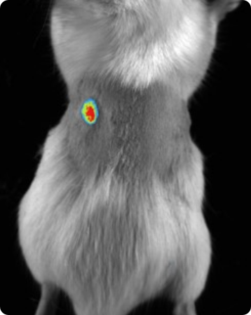
Subcutaneous tumor expressing mCherry. Image Credit: Scintica Instrumentation Inc
Bioluminescence imaging
Luciferase-expressing or secreting molecules in the target tissue can be found via bioluminescence imaging.
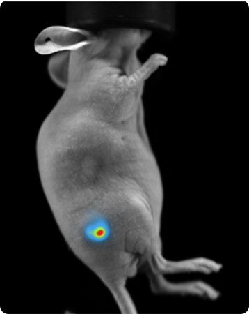
Subcutaneous tumor expressing firefly luciferase. Image Credit: Scintica Instrumentation Inc
Multispectral in vivo imaging
Different fluorescent dyes or luciferase enzyme/substrate pairings allow multispectral in vivo imaging. Up to three signals can be superimposed on the same image.
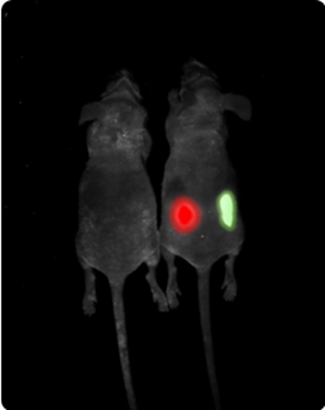
Multispectral imaging. Image Credit: Scintica Instrumentation Inc
Longitudinal imaging
- A longitudinal image sequence can be created by arranging images taken at various times. Images taken several days after an experimental therapy, for instance, could be used to create a time series.
- The image data is then compared by software throughout the experimental treatment.


Image Credit: Scintica Instrumentation Inc
Models and specifications
Newton 7.0 models BT100 and BT500
3 and 5 mice
- Platform for in vitro/in vivo optical imaging
- Three-dimensional optical tomography
- Bioluminescence detection
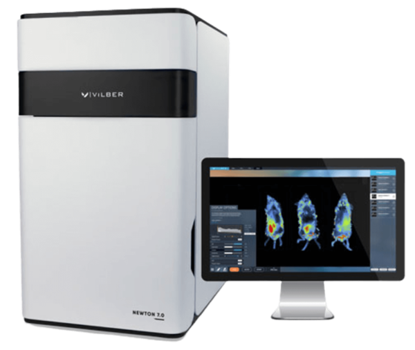
Image Credit: Scintica Instrumentation Inc
Newton 7.0 models FT100 and FT500
3, 5, and 10 mice
- Platform for in vitro/in vivo optical imaging
- Three-dimensional optical tomography
- Bioluminescence detection
- The detection of fluorescence
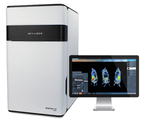
Image Credit: Scintica Instrumentation Inc
Newton 7.0 BIO plant imaging
- Platform for in vitro/in vivo optical imaging
- Fentogram for Bioluminescence Detection
- Picogram for Fluorescence Detection
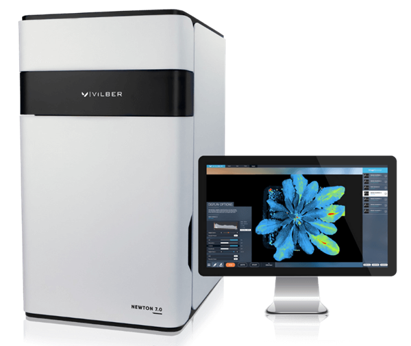
Image Credit: Scintica Instrumentation Inc
Camera - All models
- 16-bit Scientific Grade CCD Camera:
- Cooling: -90 °C absolute
- Cooling: -120 °C Delta
Lens - All models
- Fixed V.070, a proprietary version
- A motorized lens with a focal length
- Aperture: f/0.7
Resolution - All models
- Dimensions: 2160 × 2160
- Color and Monochrome Imaging
Emission - All models
- 11-All variants include a motorized filter wheel with 11 positions.
- The standard includes eight narrow band-pass filters (500, 550, 600, 650, 700, 750, 800, and 850 nm).
Excitation
Source: Scintica Instrumentation Inc.
| |
BT100 |
BT500 |
FT100 |
FT500 |
BIO |
|
White-Light
|
Dual EPI-White Light LED Panels
|
Dual EPI-White Light LED Panels
|
Dual EPI-White Light LED Panels
|
Dual EPI-White Light LED Panels
|
Dual EPI-White Light LED Panels
|
|
Fluorescence
|
Upgradeable to Fluorescence
|
Upgradeable to Fluorescence
|
8 Fluorescent Channels Included 420 / 480 / 520 / 580 / 640 / 680 / 740 / 780 nm
|
8 Fluorescent Channels Included 420 / 480 / 520 / 580 / 640 / 680 / 740 / 780 nm
|
8 Fluorescent Channels Included 420 / 480 / 520 / 580 / 640 / 680 / 740 / 780 nm
|
Emission
Source: Scintica Instrumentation Inc.
| |
BT100 |
BT500 |
FT100 |
FT500 |
BIO |
|
Filter
Wheel
|
11-position Motorized Filter Wheel
|
11-position Motorized
Filter Wheel
|
11-position Motorized Filter Wheel
|
11-position Motorized Filter Wheel
|
11-position Motorized Filter Wheel
|
|
Emission Filters
|
4 Narrow Band-pass filters included for BLI Tomography:
500/550/600/650 nm
|
4 Narrow Band-pass filters included for BLI Tomography: 500/550/600/650 nm
|
8 Narrow Band-pass filters included:
500/550/600/
650/700/750/800/850 nm
|
8 Narrow Band-pass filters included:
500/550/600/650/
700/750/800/850 nm
|
8 Narrow Band-pass filters included:
500/550/600/650/
700/750/800/850 nm
|
Darkroom
Source: Scintica Instrumentation Inc.
| |
BT100 |
BT500 |
FT100 |
FT500 |
BIO |
|
Motorization
|
- Fixed Camera
- Fixed Animal Stage
|
- Z-Axis Motorized Camera
- X/Y-Axis Motorized Animal Stage
|
- Fixed Camera
- Fixed Animal Stage
|
- Z-Axis Motorized Camera
- X/Y-Axis Motorized Animal Stage
|
- Z-axis Motorized Camera
- 15° Tilting Sample Stage
|
|
Animal Handling
|
- Heated Mouse Bed (+37 °C) included
- Animal breathers included
|
- Heated Mouse Bed (+37 °C) included
- Animal breathers included
|
- Heated Mouse Bed (+37 °C) included
- Animal breathers included
|
- Heated Mouse Bed (+37 °C) included
- Animal breathers included
|
|
Animal handling - All models
- Heated Mouse Bed (+37 °C) (included)
- Breathers for animals (inclusive)
- (Not a part of Newton 7.0 BIO.)
Accessories/add-ons
Source: Scintica Instrumentation Inc.
| |
BT100 |
BT500 |
FT100 |
FT500 |
BIO |
|
Monitorization
|
Fixed Camera
Fixed Animal Stage
|
Z-Axis Motorized Camera
X/Y-Axis Motorized Animal Stage
|
Fixed Camera
Fixed Animal Stage
|
Z-Axis Motorized Camera
X/Y-Axis Motorized Animal Stage
|
Z-axis Motorized Camera
15° Tilting Sample Stage
|
Newton 7.0 image gallery
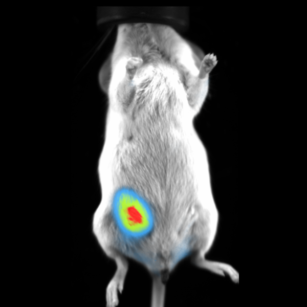
BLI Imaging of Orthotropic Mammary Fat Pad Tumor in Mouse. Image Credit: Scintica Instrumentation Inc
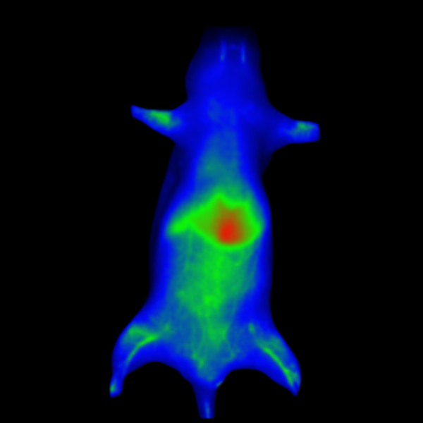
Fluorescent Nanoprimer Distribution After IV Injection in Mouse. Image Credit: Scintica Instrumentation Inc
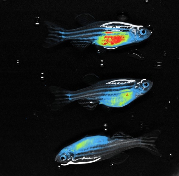
RED FLI - Zebrafish. Image Credit: Scintica Instrumentation Inc
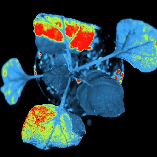
Plants- TUYV GFP Pot. Image Credit: Scintica Instrumentation Inc
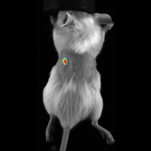
mCherry Fluorescence Imaging in Subcutaneous Tumor in Mouse. Image Credit: Scintica Instrumentation Inc
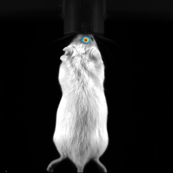
BLI Image of Orthotropic Brain Tumor in Mouse. Image Credit: Scintica Instrumentation Inc
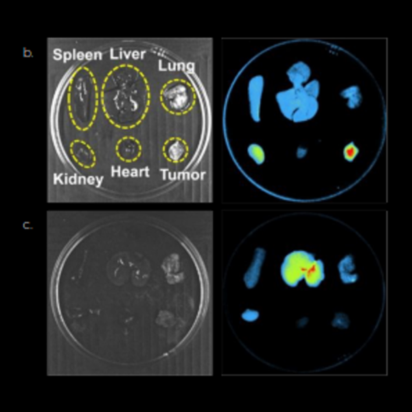
Spleen, Liver, Lung, Kidney, Heart, Tumor. Image Credit: Scintica Instrumentation Inc
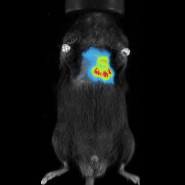
BLI Image of Orthotropic Lung Metastases in Mouse. Image Credit: Scintica Instrumentation Inc
Applications
Oncology
In preclinical animal models, optical imaging can be used to non-invasively track the development and metastasis of cancer throughout the body.
Immunology
Monitoring these populations can greatly aid in understanding the physiology of immune cell groups and creating novel treatment approaches.
Infectious disease
Optical imaging can be utilized to non-invasively visualize a site of infection and the efficacy of a treatment in the setting of a living subject.
Neurology
Optical imaging can evaluate new targeted therapies in the brain and spinal cord and track the development of different neurodegenerative disorders.
Biodistribution studies
In preclinical biodistribution research, optical imaging offers a distinct benefit due to its ability to view the entire body; a single image can measure several organs throughout the body.