For real-time, in vivo screening of all single photon emission computed tomography (SPECT) isotopes, the γ-eye is the first and only SPECT scanner. The γ-eye is a comprehensive solution for all labs at any research stage because of its interchangeable collimators, compact size, and field of vision appropriate for full-body mouse imaging.
The system weighs less than 40 kg and measures only 44 cm by 46 cm by 40 cm. It is a desktop solution that can transform any area into an image lab. The γ-eyes have a sensitivity of 341 cps/MBq, a spatial resolution of up to 1.9 mm, and an energy resolution of less than 19%, all while having a compact footprint.
The Visual|eyes software suite offers an integrated, user-friendly platform for picture acquisition and data analysis, while the animal handling system guarantees the health and welfare of the imaging subject.
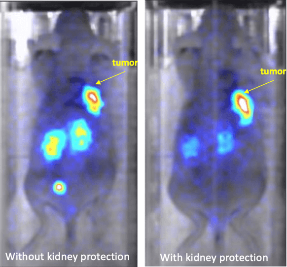
Image Credit: Scintica Instrumentation Inc.
Features and benefits
Realtime imaging from time zero post-injection
If the injection was effective and the radioisotope could be imaged inside the imaging subject, the user would receive an immediate response.
Frame rates down to 1 second
When coupled with the previously mentioned features of real-time, whole-body, and mouse imaging, these acquisition periods would enable tracking the imaging compound's kinetics and dynamic distribution throughout time.
Small footprint (44 cm X 46 cm X 40 cm), and weight less than 40 kg
The system is genuinely tabletop due to its weight and tiny size.
Easy-to-use analysis software and export capabilities, including DICOM filed
The analysis program allows users to rapidly and simply generate quantitative data on their established ROIs. Alternatively, users can export the data and utilize third-party applications to analyze it.
Animal handling system with integrated anesthesia delivery, heated bed, and option to monitor vital signs
Throughout the session, the user can monitor and steady the imaging subject's physiological condition. This is crucial for the animal's health and welfare and enables consistency in data.
Active field of view of 50 mm X 100 mm
This field of view enables continuous and dynamic imaging in a single acquisition, making it appropriate for whole-body mouse imaging. Examining the imaging compound's kinetics and its temporal and spatial dispersion calls for this.
State-of-the-art technical characteristics
- cps/MBq sensitivity of 341
- Resolution of space up to 1.9 mm
- Less than 19% energy resolution
- Dynamic range between 30 and 500 keV
- Collimators that can be swapped out
Offers high-sensitivity images over a wide range of signal intensities over the entire field of view.
Simple connections
- Electrical requirements: 100-240 VAC
- USB 2.0 Type A and Gb Ethernet PC connectivity
Permit the system to be installed in any clean room, laboratory, or animal facility to transform it into an imaging lab.
Anatomical mapping using artificially generated X-ray images
Since the system lacks an X-ray source, it does not emit radiation; yet, the resulting X-ray image still provides anatomical information.
Common imaging Bed for all eye systems
When an animal has a shared imaging bed, it can travel from one ocular system to another without moving. The ability to co-register images makes multimodal imaging possible.
Models and specifications
Performance
- cps/MBq sensitivity 341
- Resolution of space up to 1.9 mm at 0 mm
- Less than 19% energy resolution
- Dynamic range between 30 and 500 keV
- Time intervals, as short as one second
Animal handling
- Integrated anesthesia
- The animal stage that is heated
- Monitoring of vital signs
Image Credit: Scintica Instrumentation Inc.
Tech specs
- Photomultiplier tubes or photodetectors
- CsI: Na Scintillators
- Field of view active = 50 × 100 mm
- Anatomical mapping using synthetic X-rays
- General-purpose, high resolution, high sensitivity, and high energy interchangeable collimators.
γ-eye imaging gallery

SPECT imaging of IN-111-Dotatate in mice with tumors. Image on the right includes protection of the kidneys, as seen by the reduced signal in this organ. Image Credit: Scintica Instrumentation Inc.
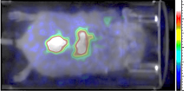
This image shows Tc-99 perfusion imaging in a mouse. This image was acquired in real-time with 1 second frame rate. Image Credit: Scintica Instrumentation Inc.
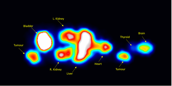
Mouse phantom filled with Tb-161 imaged with General Purpose collimator. Image Credit: Scintica Instrumentation Inc.
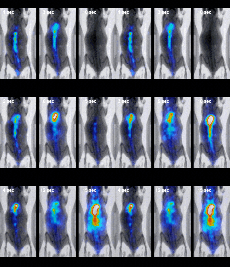
Image Credit: Scintica Instrumentation Inc.
Applications
Cancer research
SPECT can be applied to cancer research in numerous ways: It can verify whether malignancies are present, track the size and progression of tumors, identify metastases, and determine whether particular biomarkers are expressed.

SPECT imaging of IN-111-Dotatate in mice with tumors. Image on the right includes protection of the kidneys, as seen by the reduced signal in this organ. Image Credit: Scintica Instrumentation Inc.
Biomarker detection
Certain biomarkers can be imaged using targeted probes, such as:
- Inflammation
- Hypoxia
- Angiogenesis
- etc.
Theranostics
Numerous SPECT compounds thought to be simultaneously therapeutic and diagnostic are being produced.
Pharmacokinetics/dynamics/biodistribution
Following injection to the imaging patient, labeled compounds can be utilized to track the compound's pharmacokinetics, pharmacodynamics, and biodistribution over time.