Curiosis has a line of Automated Live Cell Imaging Systems called the Celloger series. Using specialized microscopes and labeling procedures, live cell imaging enables real-time observation and investigation of biological processes within living cells or organisms.
The live-cell imaging approach employs time-lapse microscopy to observe the intricate dynamics of living cells in real-time, allowing for the comprehension and investigation of a wide range of biological processes.
Real-time imaging of biological processes such as cell migration, development, and trafficking greatly benefits research in cell biology, neuroscience, pharmacology, and developmental biology.
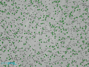
Image Credit: Scintica Instrumentation Inc.
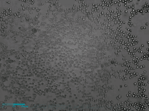
Image Credit: Scintica Instrumentation Inc.
Founded in 2015, Curiosis is headquartered in Korea. The business creates, manufactures, and distributes life science lab equipment. Curiosis offers a way to increase the effectiveness of cell-based research by creating several products for precise cell analysis, which is the foundation of bioscience research.
Numerous cell research procedures, including cell imaging, cell separation, and cell counting, involve Curiosis products. The groundbreaking Live Cell Imaging line, known as the Celloger series, redefines research capabilities.
With its unparalleled ease of use and outstanding image quality, it gives researchers access to cutting-edge technologies that facilitate real-time cell monitoring within the incubator. This allows for the smooth tracking and observation of cellular dynamics without interfering with the natural growing environment.
Long-term imaging is made possible by the Celloger series' ability to withstand self-generated heat and their small size, which allows them to be kept in a standard cell culture incubator.
Furthermore, optimizing the fluorescence filter and light path can provide fluorescence images of live cells in real-time with the least light intensity and clear bright-field images utilizing contrast-enhanced optics.
Features and benefits
Real-time cell monitoring inside an incubator
The Celloger series is made to track cells effectively in real time without interfering with the circumstances necessary for cell growth. Researchers can remotely view cells in real-time by setting up the devices within the incubator and connecting them to an external PC.
Time-lapse imaging capability
Cell photos are automatically taken using the time-lapse feature by the researcher's timetable, and the photographs can be quickly transformed into time-lapse videos.
Compatible with different vessels types
To accommodate a wide range of experiments, different cell culture vessels such as well plates (up to 96 wells), flasks, dishes, and slides can be used by simply replacing the vessel holders for specific needs. Celloger Stack is used for multi-layer vessel types.
User-friendly software
Users can make infinite copies of the Scanning and Analysis software since they are offered as standard packages. With the help of various analysis tools included in these programs, researchers may quickly configure numerous image capture modes and produce useful experimental data.
Models
Celloger pro
- Fluorescence in several colors and brightfield imaging
- Monitoring in real-time within the incubator
- Objective lens that the user can change
- Taking pictures from various angles
- Suitable for a variety of vessel types
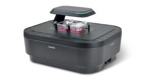
Image Credit: Scintica Instrumentation Inc.
Celloger stack
- Monitoring in real-time within an incubator
- Indicates when to harvest cells
- The interchangeable vessel holders can be used with a variety of vessels
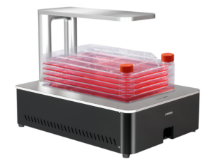
Image Credit: Scintica Instrumentation Inc.
Celloger nano
- Various kinds of vessels
- Adapt to standard CO2 incubators
- Provide user-friendly features
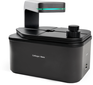
Image Credit: Scintica Instrumentation Inc.
Celloger mini-plus
- Monitoring in real time
- Small size
- Imaging from many points
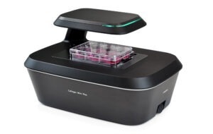
Image Credit: Scintica Instrumentation Inc.
Cellpuri
- Cell enrichment
- Filterless Filter (FLF) technology
- Decreased clumping of cells
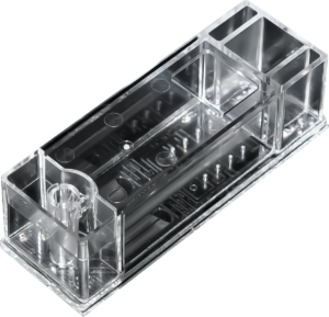
Image Credit: Scintica Instrumentation Inc.
Technical specifications
Source: Scintica Instrumentation Inc.
| Features |
Celloger NANO |
Celloger MINI-PLUS |
Celloger PRO |
Celloger STACK |
| APPLICATION |
Live Cell Imaging |
Live Cell Imaging |
Live Cell Imaging |
Live Cell Imaging |
| IMAGE MODE |
Bright-field,
Single color
Fluorescence
(Green/Red) |
Bright-field,
Single color
Fluorescence
(Green/Red) |
Bright-field,
Dual color
Fluorescence
(Green/Red) |
Bright-field |
| Green |
Ex: 470/40 Em: 510lp |
Ex: 470/40 Em: 540/50 |
Ex: 470/40 Em: 510lp |
|
| Red |
Ex: 525/30 Em: 570lp |
Ex: 525/30 Em: 570lp |
Ex: 562/40 Em: 641/75 |
|
| Camera |
1.25 MP CMOS |
5 MP CMOS |
Ex: 562/40 Em: 641/75 |
5 MP CMOS |
| Position |
Single |
Multiple |
Multiple |
Multiple |
| Stage type |
Manual XY, automatic Z
moving |
Automatic XYZ moving
(motorized camera) |
Automatic XYZ moving
(motorized camera) |
Automatic XYZ moving
(motorized camera) |
| Focusing |
Autofocusing & Manual
focusing |
Autofocusing & Manual
focusing |
Autofocusing & Manual
focusing |
Autofocusing & Manual
focusing |
| Objective Lens |
2X/ 4X / 10X |
2X/ 4X / 10X |
2X/ 4X / 10X
(User-Interchangeable) |
2X |
Vessels Holder
Compatibility |
96 well plate, flasks,
dishes and slides |
96 well plate, flasks,
dishes and slides |
96 well plate, flasks,
dishes and slides |
Multi-layer vessel types |
| Time-Lapse Imaging |
Yes |
Yes |
Yes |
Yes |
Scanning and Analysis
Software |
Included |
Included |
Included |
Included |
| Operating Environment |
10-40 ℃, 20-95% humidity |
10-40 ℃, 20-95% humidity |
10-40 ℃, 20-95% humidity |
10-40 ℃, 20-95% humidity |
| Dimension |
211 x 146 x 188 mm |
226 x 358 x 215 mm |
250 x 338 x 412 mm |
250 x 338 x 412 mm |
| Dimension |
3.2 Kg |
5.6 Kg |
9.6 Kg |
15 Kg |
Applications
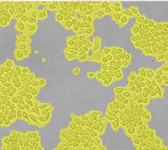
Cell Proliferation. Image Credit: Scintica Instrumentation Inc.
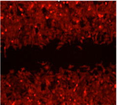
Wound-healing. Image Credit: Scintica Instrumentation Inc.
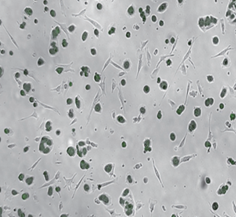
Co-culture monitoring. Image Credit: Scintica Instrumentation Inc.
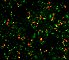
Transfection Efficiency. Image Credit: Scintica Instrumentation Inc.
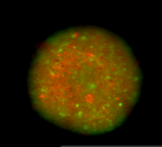
Spheroid Cytotoxicity. Image Credit: Scintica Instrumentation Inc.
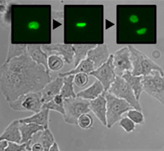
Cell Monitoring. Image Credit: Scintica Instrumentation Inc.
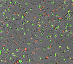
Apoptosis and Cytotoxicity. Image Credit: Scintica Instrumentation Inc.
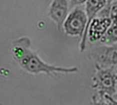
Phagocytosis. Image Credit: Scintica Instrumentation Inc.
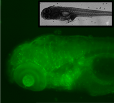
Zebra Fish Observation. Image Credit: Scintica Instrumentation Inc.
Accessories and add-ons
Source: Scintica Instrumentation Inc.
| Part number |
Description |
| CUR-CRCLG-MPWPS |
Vessel holder Well plate 96 |
| CUR-CRCLG-MPTFS25 |
Vessel holder A25 cm2 single |
| CUR-CRCLG-MPTFD25 |
Vessel holder A25 cm2 (Dual) |
| CUR-CRCLG-MPTFS75 |
Vessel holder A75 cm2 (Single) |
| CUR-CRCLG-MPPDD35 |
Vessel holder Petri dish 35 mm |
| CUR-CRCLG-MPPDD60 |
Vessel holder Petri dish 60 mm |
| CUR-CRCLG-MPPDS90 |
Vessel holder Petri dish 90/100 mm |
| CUR-CRCLG-MPSH03 |
Vessel holder slide |
| CUR-CRCPR-SP01 |
Syringe pump (Motorized vertical) |
| CUR-CRCSD-NI50 |
C-slide – Disposable Hemocytometer (Neubauer Improved) 50 slides |
Video resources - Celloger series
Celloger® Nano, automated live cell imaging system
Celloger® Nano, automated live cell imaging system. Video Credit: Scintica Instrumentation Inc.
Celloger® Mini Plus, automated live cell imaging system
Celloger® Mini plus, automated live cell imaging system. Video Credit: Scintica Instrumentation Inc.
Celloger® Pro, automated live cell imaging system
Celloger® Pro, automated live cell imaging system. Video Credit: Scintica Instrumentation Inc.