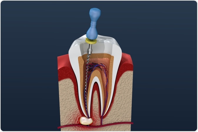
In the field of endodontics, clinicians are often tasked with trying to remove several species of harmful microorganisms that affect the root canal, many of which can cause serious damage if left unattended.1
Reports suggest there is a 75% success rate for root canal treatment.2 Although high, some of these cases are associated with post-treatment disease. While cleaning and shaping of the root canal are essential components of endodontics, the root canal system contains many anatomical complexities that prevent successful cleaning and endodontic treatment.
 Image Credit: Alex Mit / Shutterstock.com
Across the population, there is variation in canal shape, with some people having two or more canals in one root. Additionally, current preparation and irrigation techniques possess inherent limitations that can reduce the successful eradication of these microorganisms from the root canal system. Even the most effective techniques for complete disinfection, whether chemical or mechanical, are less than ideal when faced with the curvatures of narrow canals.
The first option of choice for post-endodontic disease is nonsurgical root canal treatment, with retreatment consisting of complete removal of the dental materials in the root canal system. Inflammatory and pain-causing necrotic tissue or bacteria is sometimes found among the filling material, necessitating prompt removal.
Stainless steel (SS) hand files, nickel-titanium (Ni-Ti) rotary instruments, and ultrasonic tips represent some of the techniques used for removing root canal filling material.
Unfortunately, none of the available retreatment strategies can clean the root canal wall completely, especially in the apical third.3,4 Since many instruments are limited by the anatomical structure within the canal system, it is essential for operators to select an instrument that can clean the Gutta–Percha (GP) cone and sealer debris in the apical third effectively.
Brush system superior to hand files and ultrasound irrigation
In a 2019 study, researchers from Korea examined the efficacy of three different canal filling material removal techniques after filling with a Gutta–Percha cone and calcium silicate-based sealer in artificial teeth.5 These three techniques included hand files, passive ultrasonic irrigation, and brush irrigation.
According to the researchers, the brush irrigation technique can aid retreatment by removing Gutta–Percha cone and calcium silicate-based sealer particles that become stuck on the canal walls.
The study investigators measured the percentage of volume debris of Gutta–Percha and sealer that remained within the canal using the SkyScan 1272, a high-resolution micro computed tomography device.6
This device offers nondestructive imaging of up to 209 Megapixel virtual slices through objects as small as 0.35um. Objects as small as this can be detected due to the platform’s phase-contrast enhancement.
The SkyScan was applied to samples with each of the three techniques to render three-dimensional (3D) images of the filling material by surface-CT-Vol. Additionally, the instruments’ capabilities allowed researchers to quantify the volume of the prepared canal space with each of the sample specimens while evaluating the volume of residual filling material following retreatment using the accompanying CT-An software.
Investigators of this study defined the apical region as the area between 1 mm and 5 mm from the apex.
Overall, the researchers found that the brush system was superior in the cleaning of the root canal compared to the retreatment files and the passive ultrasonic irrigation system in the apical area. In the ultrasonic irrigation group, the majority of residual material was found in the apical region following retreatment.
Study findings
According to the CT scans, the total average post-retreatment volumes of residual filling were 4.84896 mm3, 0.80702 mm3, and 0.05248 mm3 for the conventional file, ultrasound irrigation, and brush irrigation groups, respectively.
Additionally, the group that received file cleaning had a remaining 6.76% of filling material within the canal versus 1.12% in the passive ultrasound irrigation group and 0.07% in the brush irrigation group.
Limitations and possible clinical implications
Compared to conventional retreatment files, the researchers argued, additional passive ultrasound irrigation can eliminate sealer and Gutta–Percha debris extending into areas that can’t be reached.
The brush irrigation technique may be superior to both ultrasound irrigation and conventional files, based on the study’s findings, and the researchers suggest recommending this technique whenever feasibly possible.
The benefits of the brush technique may be due to the form of the brush strands, which twist when the brush is not rotating and open when the device is rotating at high speeds. When the device is rotating, the strands cover the entire diameter of the canal, removing remaining debris in a mechanical fashion. Additionally, the strands bring the irrigant into contact with the surface of the canal.
Certain limitations of this study may prevent the generalizability of these findings across current clinical practice. Artificial teeth in this study, for instance, aren’t entirely identical to natural human teeth, as there is often variation and substantially more complexity in root canal anatomy.
Despite this and other limitations inherent in this study, the SkyScan system was able to demonstrate the superiority of a brush system over conventional root canal retreatment cleaning strategies, due to the tool’s flexibility across the canal’s curvature. Whether or not this finding can be replicated in human patients requires further study.
References:
- Peciuliene V, Maneliene R, Balcikonyte E, Drukteinis S, Rutkunas V. Microorganisms in root canal infections: a review. Stomatologija. 2008;10(1):4-9.
- Ng YL1, Mann V, Rahbaran S, Lewsey J, Gulabivala K. Outcome of primary root canal treatment: systematic review of the literature - part 1. Effects of study characteristics on probability of success. Int Endod J. 2007;40(12):921-39.
- Giuliani V, Cocchetti R, Pagavino G. Efficacy of ProTaper universal retreatment files in removing filling materials during root canal retreatment. J Endod. 2008;34(11):1381-1384.
- Mollo A, Botti G, Prinicipi Goldoni N, et al. Efficacy of two Ni-Ti systems and hand files for removing gutta-percha from root canals. Int Endod J. 2012;45(1):1-6.
- Nguyen TA, Kim Y, Kim E, Shin SJ, Kim S. Comparison of the efficacy of different techniques for the removal of root canal filling material in artificial teeth: a micro-computed tomography study. J Clin Med. 2019 Jul 7;8(7).
- Bruker SkyScan. Accessed February 10, 2020.
About Bruker BioSpin Group
The Bruker BioSpin Group designs, manufactures, and distributes advanced scientific instruments based on magnetic resonance and preclinical imaging technologies. These include our industry-leading NMR and EPR spectrometers, as well as imaging systems utilizing MRI, PET, SPECT, CT, Optical and MPI modalities. The Group also offers integrated software solutions and automation tools to support digital transformation across research and quality control environments.
Bruker BioSpin’s customers in academic, government, industrial, and pharmaceutical sectors rely on these technologies to gain detailed insights into molecular structure, dynamics, and interactions. Our solutions play a key role in structural biology, drug discovery, disease research, metabolomics, and advanced materials analysis. Recent investments in lab automation, optical imaging, and contract research services further strengthen our ability to support evolving customer needs and enable scientific innovation.
Sponsored Content Policy: News-Medical.net publishes articles and related content that may be derived from sources where we have existing commercial relationships, provided such content adds value to the core editorial ethos of News-Medical.Net which is to educate and inform site visitors interested in medical research, science, medical devices and treatments.