The biological importance of intrinsically disordered proteins (IDPs) has been discovered in recent years. Sequence-specific assignments using NMR, while well developed for globular proteins, are more challenging for IDPs. This article outlines some of the newly developed NMR techniques for detecting amino acid residues in IDPs with spatial resolution.
 Image Credit: Naeblys / Shutterstock.com
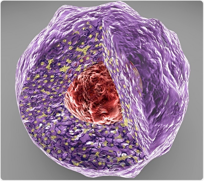
Intrinsically disordered proteins (IDP) are proteins that lack tertiary structure. They are now being recognized as biologically crucial in both healthy systems and the pathophysiology of various diseases. Novel therapies could target relevant IDPs, but to do this, we need to fully understand their structures and how they function under their native physiological conditions.1
Due to their lack of structure and high mobility, studying IDPs can be challenging. They cannot be characterized using traditional X-ray crystallography techniques, so nuclear magnetic resonance (NMR) is the tool of choice. NMR makes it relatively easy to identify IDPs, yet studying their structure and dynamics raises several challenges. Fortunately, there are an increasing number of NMR tools to overcome these difficulties.2,3
Assigning Proline Residues in IDP NMR
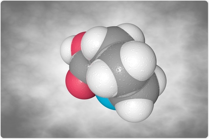
Proline plays an essential role in the structure of many IDPs because its cyclic structure and lack of hydrogen bonding can prevent the formation of secondary structures.
Detecting proline is vital to understanding the structure and dynamics of IDPs. However, proline signals are usually invisible in 1H-15N NMR experiments because they lack a hydrogen atom on their alpha-amino group.4
A 2018 publication from researchers at the University of Florence, Italy, in ChemBioChem explains how they used 13C direct‐detection NMR spectroscopy to detect proline residues in IDPs.
The researchers used 2D CON, which correlates the amide nitrogens of a residue with the 13C spin in the proceeding residue. Their NMR spectra were acquired on a Bruker AVANCE NEO spectrometer equipped with a cryogenically cooled probe head optimized for 13C-direct detection (TXO).4
Using this technique, the scientists were able to resolve signals from different proline residues in an IDP. Using band-selective 180° pulses on 15N spins, they exploited the unusual 15N shifts of proline to produce selective proline fingerprint spectra. They obtained a fingerprint spectrum for an IDP called CBP-ID4 and successfully detected all 45 proline residues in the protein, with almost no spectral overlap.4
This simple technique will undoubtedly enhance the study of IDP dynamics and structure using NMR, which are frequently reported without proline residues.
 Image Credit: Maryna Olyak / Shutterstock.com
Overcoming low chemical shift dispersion
Assignment of NMR signals is typically achievable for globular proteins using 3D triple-resonance experiments to find neighboring backbone atoms, relying on coherence transfer steps involving 13C, 15N, and 1H.3,5
However, high flexibility and a solvent-exposed backbone can impact NMR spectra, resulting in low chemical shift dispersion and noise. This can make it challenging to resolve signals, and almost impossible to assign signals.3,5
NMR spectroscopy, using heteronuclear spins, is characterized by a large chemical shift dispersion, which can be exploited to characterize IDPs. However, 3D triple resonance does not provide the necessary resolution to enable complete signal assignment in complex IDPs. High resolution in multi-dimensions is required.3,5,6
Today, direct detection of heteronuclei (13C) using cryoprobe technology, non-uniform sampling for high-dimension experiments, and fa
Complete assignment of IDPs with projection spectroscopy
st acquisition provide the necessary resolution to overcome the challenges caused by low chemical shift dispersion in IDPs. As a result, we are beginning to see the complete assignment of IDPs with NMR.3,5
A second publication by the team from the University of Florence outlines how they used projection spectroscopy in 13C-direct detected NMR experiments to achieve the sequence-specific assignment of IDPs.
The study, which was published in the Journal of Biomolecular NMR in 2018, outlines how they used the technique to perform the complete assignment of alpha-synuclein signals, a biologically interesting IDP that has been linked to Parkinson's disease and other neurodegenerative diseases.
Automated projection spectroscopy (APSY) is a technique that involves obtaining discrete sets of projection spectra in high-dimension NMR experiments, combined with automatic identification of chemical shift correlations using an algorithm called GAPRO (geometric analysis of projections).
GARPO identifies peaks that arise from the same resonance and calculates their positions in the N-dimensional frequency space. APSY is fully automated and produces high precision chemical shift measurements, while eliminating noise, enabling complete sequence assignment of IDPs.6
The Italian research team conducted a series of NMR experiments on 13C, 15N labeled human alpha-synuclein, collecting multidimensional spectra. All their 2D and APSY NMR experiments were conducted using a Bruker AVANCE NEO spectrometer equipped with a cryogenically cooled probe head optimized for 13C-direct detection (TXO).
Using their technique, they obtained excellent resolution chemical shift measurements. They were able to assign the chemical shifts of C' and N atoms in peptide bonds along the backbone of alpha-synuclein, obtaining information on the kind of amino acid present at each position on the backbone.6
Detection of post-translational modifications
IDPs, like all proteins, can be subjected to post-translational modifications. Such changes are often essential for protein function and can be crucial in disease mechanisms. It is for this reason that they must be reproduced when synthesizing IDPs for research and analysis. However, replicating post-translational modifications and studying them using NMR can be difficult.7
A recent paper by scientists from Masaryk University in the Czech Republic describes how they produced IDPs that were phosphorylated after translation. They used 1D 31P NMR to detect the presence of phosphorylation, and 2D 1H-13C, 1H-15N HSQC experiments to confirm the phosphorylated residues. Their NMR spectra were obtained with a Bruker AVANCE III HD spectrometer with a QCI cryoprobe for 31P experiments, and a TXI probe for all the other experiments. 7
Their 31P spectra successfully confirmed two phosphorylations in their model peptide, while HSQC experiments identified tyrosine as the phosphorylated residue. Research into replicating and detecting other post-transitional modifications in IDPs is still on-going. 7
Summary
In recent years, NMR has evolved into a powerful technique in structural biology for studying the dynamics of proteins, including IDPs. It is now possible to overcome the problems of incomplete assignment, low chemical shift dispersion, and an inability to detect post-translational modifications using new techniques.
Applying these techniques to biologically relevant IDPs may open the door for new therapies targeting a range of diseases.
References
- ‘Structure and Function of Intrinsically Disordered Proteins’ - Tompa P, Fersht A, CRC, 2009.
- ‘Application of NMR to studies of intrinsically disordered proteins’ - Gibbs EB, Cook EC, Showalter SA, Archives of Biochemistry and Biophysics, 2017.
- ‘NMR contributions to structural dynamics studies of intrinsically disordered proteins’ - Konrat R, Journal of Magnetic Resonance, 2014.
- ‘Prolines’ fingerprint in intrinsically disordered proteins’ - Murrali MG, Piai A, Bermel W, Felli IC, Pierattelli R, ChemBioChem, 2018.
- ‘Intrinsically Disordered Proteins Studied by NMR Spectroscopy’ - Felli IC, Pierattelli R, Springer, 2015.
- ‘13C APSY-NMR for sequential assignment of intrinsically disordered proteins’ - Murrali MG, Schiavina M, Sainati V, Bermel W, Pierattelli R, Felli IC, Journal of Biomolecular NMR, 2018.
- ‘Efficient and robust preparation of tyrosine phosphorylated intrinsically disordered proteins’ - Brázda P, Sedo O, Kubícek K, Štefl R, BioTechniques, 2019.
About Bruker BioSpin Group
The Bruker BioSpin Group designs, manufactures, and distributes advanced scientific instruments based on magnetic resonance and preclinical imaging technologies. These include our industry-leading NMR and EPR spectrometers, as well as imaging systems utilizing MRI, PET, SPECT, CT, Optical and MPI modalities. The Group also offers integrated software solutions and automation tools to support digital transformation across research and quality control environments.
Bruker BioSpin’s customers in academic, government, industrial, and pharmaceutical sectors rely on these technologies to gain detailed insights into molecular structure, dynamics, and interactions. Our solutions play a key role in structural biology, drug discovery, disease research, metabolomics, and advanced materials analysis. Recent investments in lab automation, optical imaging, and contract research services further strengthen our ability to support evolving customer needs and enable scientific innovation.
Sponsored Content Policy: News-Medical.net publishes articles and related content that may be derived from sources where we have existing commercial relationships, provided such content adds value to the core editorial ethos of News-Medical.Net which is to educate and inform site visitors interested in medical research, science, medical devices and treatments.