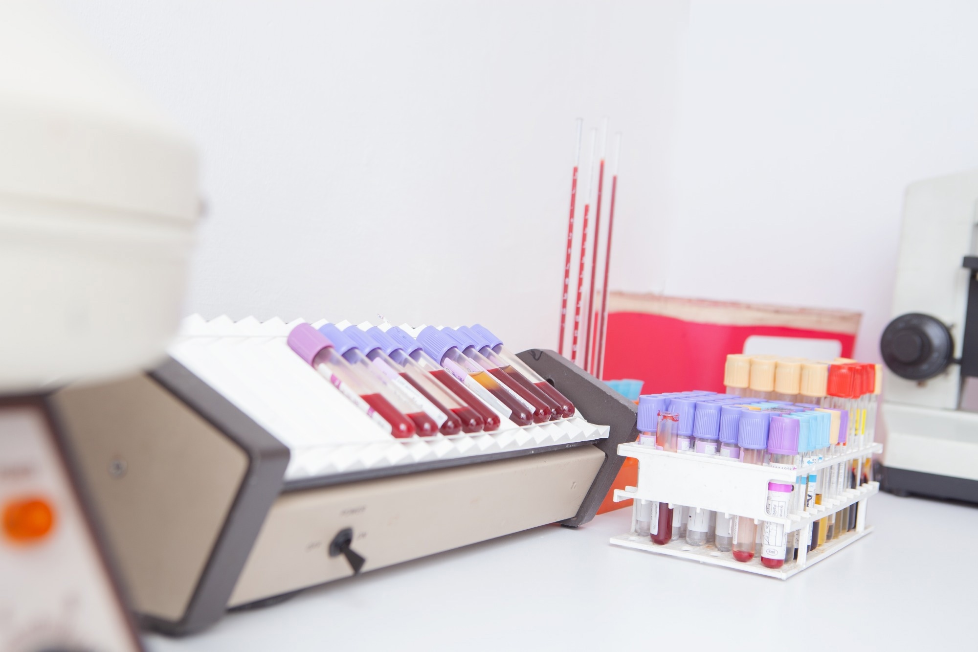
Tissue biospecimens (TBs) are advantageous for profiling genetic disorders and are essential in developing molecular biology within contemporary medicine.
Human TBs have aided the modern development of precision medicine as they allow diverse therapeutics to be tested against a range of cancer tissue subtypes while advising on the variations between samples at a molecular level.
This paper focuses on how TBs support the contemporary development of antibody–drug conjugates (ADCs) and examines the importance of Ki67 as a quality marker.

Image Credit: Yuls26/Shutterstock
Significance of tissue biospecimens
TBs have previously been utilized to classify particular molecular therapies against the estrogen and progesterone receptors and HER2 (typical biomarkers against breast cancer).
Through the use of breast cancer tissue specimens, researchers have been able to identify that HER2 overexpression is critical in the development of an aggressive breast cancer subtype in 15 to 20 % of breast cancer patients, leading to the development of Herceptin as an auxiliary drug for chemotherapy.
Classical chemotherapy treatments are aggressive and destructive at the global level. New methods require a more direct approach for localizing therapy to the disorder site while minimizing non-target cells.
Researchers have focussed on the molecular level to contest the absence of selectivity in such therapies, leading to the contemporary popularity of ADCs as potential cancer therapies.
Branded as a “smart-chemotherapy”, ADCs’ purpose is to transport chemotherapeutic compounds to malignant tissue with high efficiency. ADCs amalgamate the selectivity of targeted therapies with the cytotoxic properties of chemotherapeutic agents, representing the future of chemotherapy research.
What are ADCs?
ADCs can be divided into three chief components: the antibody, a linker, and a cytotoxic active component, which link cytotoxic treatment specifically to the region of interest.
The ideal target is especially expressed in high levels on the tumor cell's surface, reducing toxicity by decreasing the radii of cytotoxic chemical exposure. The linker fuses the active and cytotoxic components to an antibody, which will be directed to the biomarker of interest.
As the linker is steady within the bloodstream, the cytotoxic compound is present in the malignant tissue in increased concentrations, suggesting a more potent response.
Significantly, the linker component can be manufactured to be cleavable at the molecular pathology site or during antibody recognition and internalization, enabling therapy development to suit treatment against a specific cancer subtype.
By controlling the site of linker cleavage, scientists can enhance efficacy for a more measured response. After the linker cleavage, the cytotoxic payload is released at the site of interest, bringing about the localized decay of the malignant tissue.
Why ADCs are important
The use of ADCs enables the use of more potent compounds during cancer therapy; systemic administration using standard procedure would cause significant damage.
Their efficacy as chemotherapeutics depends on the dosage and linker properties: each works to regulate the payload concentration at the malignant cell surface. By using ADCs against a cancer cell, a more active component can be distributed with higher specificity and better responsiveness.
ADCs are now the gold standard in treating HER2, HR, and triple-negative breast cancer subtypes due to their active response and high specificity. Their prominence in breast cancer treatment is a result of well-characterized surface molecular biology.
Comprehension of surface microbiology is vital in developing ADCs because the antibody must be targeted to a protein on the tumor cell surface specific to the cancer tissue subtype.
If the ADC is transported to other body regions, there is a risk of substantial toxic side effects due to the potent chemotherapeutics used in the conjugates.
Ki67 and high-quality ADC generation
High-quality tissue specimens must be used during development to reduce negative side effects. Charting surface protein localization will enlighten researchers on the optimal surface antigens for targeting ADCs.
High-quality tumor tissue samples are crucial because biospecimens are used throughout ADC development, primarily within tumor biology, target validation, and ADC efficacy testing stages. Using such samples will guarantee that the ADCs produced have the maximum accuracy conceivable while maintaining specificity to curb possible side effects.
Ki67, a surface protein expressed in actively dividing cells and repressed in resting cells, is a primary marker for TB quality. Ki67 is frequently used in laboratories because its surface concentration and expression patterns are consistent.
Staining against Ki67 makes it possible to identify tumorigenic regions within a block of tissue, validating perturbed proliferation in the sample.
Ki67 is linked with aggressive disorder and a poorer prognosis and hence can act as a marker for high-risk tumors. It can be used with further immunohistochemical markers to validate target antigen expression.
By comparing the target antigen expression with Ki67, researchers can ascertain which patients would benefit from potential ADC development and identify the specificity of surface antigens by overlapping with stains against Ki67. This is valuable for ADC development, as treatment can be tailored to stages of malignancy by developing antigens against a range of biomarkers.
By integrating Ki67 block staining into the research protocol, researchers can demonstrate that their ADC research is founded on high-quality TB backgrounds.
To aid researchers in ADC development, BioIVT provides Ki67 staining procedures with pre-stained blocks and custom screens. By offering stained slide images, BioIVT accelerates research by supporting researchers in selecting between a range of tissue blocks.
The established nature of Ki67 in both research and clinical settings makes analysis simple, with translatable results that will aid research within and between laboratories.
References and further reading
- Krzyszczyk, P. et al. (2018). The growing role of precision and personalized medicine for cancer treatment. Technology (Singap World Sci.) Available at: https://www.ncbi.nlm.nih.gov/pmc/articles/PMC6352312/
- Al Diffalha, S. et al. (2019). The Importance of Human Tissue Bioresources in Advancing Biomedical Research. Biopreserv Biobank. Available at: https://www.ncbi.nlm.nih.gov/pmc/articles/PMC7061295/
- Tarantino, P. et al. (2022). Antibody-drug conjugates: Smart chemotherapy delivery across tumor histologies. CA Cancer J Clin. Available at: https://pubmed.ncbi.nlm.nih.gov/34767258/
- Shastry, M. et al. (2023). Rise of Antibody-Drug Conjugates: The Present and Future. Am Soc Clin Oncol Educ Book Available at: https://pubmed.ncbi.nlm.nih.gov/37229614/
- Fu, Z. et al. (2022). Antibody drug conjugate: the “biological missile” for targeted cancer therapy Sig Transduct Target Ther Available at: https://www.nature.com/articles/s41392-022-00947-7#citeas
- Sun, X., Kaufman, PD. (2018). Ki-67: more than a proliferation marker. Chromosoma. Available at: https://www.ncbi.nlm.nih.gov/pmc/articles/PMC5945335/
- Miller, I. et al. (2018). Ki67 is a Graded Rather than a Binary Marker of Proliferation versus Quiescence. Cell Rep. Available at: https://www.ncbi.nlm.nih.gov/pmc/articles/PMC6108547/#:~:text=Ki67%20staining%20is%20frequently%20used,a%20percent%20Ki67%2Dpositive%20value.
- BioIVT. (2024). Assessing the Research Value of Biospecimens for ADC development with KI67 tissue staining. [Online] BioIVT. Available at: https://bioivt.com/blogs/assessing-the-research-value-of-biospecimens-for-adc-development-with-ki67-tissue-staining
About BioIVT
BioIVT, formerly BioreclamationIVT, is a leading global provider of high-quality biological specimens and value-added services. We specialize in control and disease state samples including human and animal tissues, cell products, blood and other biofluids. Our unmatched portfolio of clinical specimens directly supports precision medicine research and the effort to improve patient outcomes by coupling comprehensive clinical data with donor samples.
Our Research Services team works collaboratively with clients to provide in vitro hepatic modeling solutions. And as the world’s premier supplier of ADME-Tox model systems, including hepatocytes and subcellular fractions, BioIVT enables scientists to better understand the pharmacokinetics and drug metabolism of newly discovered compounds and the effects on disease processes. By combining our technical expertise, exceptional customer service, and unparalleled access to biological specimens, BioIVT serves the research community as a trusted partner in ELEVATING SCIENCE®.
Sponsored Content Policy: News-Medical.net publishes articles and related content that may be derived from sources where we have existing commercial relationships, provided such content adds value to the core editorial ethos of News-Medical.Net which is to educate and inform site visitors interested in medical research, science, medical devices and treatments.