Live cell imaging is a technique for studying living cells over time utilizing images captured by time-lapse microscopy.
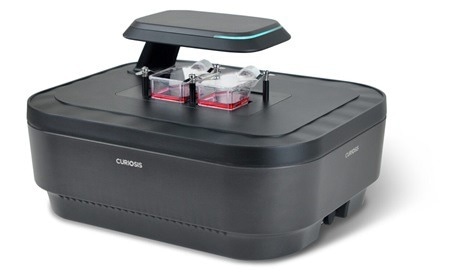
Celloger. Image Credit: Scintica Instrumentation Inc.
Real-time imaging of cellular processes such as cell migration, development, and trafficking can advance research in various academic domains, including cell biology, cancer research, neuroscience, pharmacology, and developmental biology.
To see the cells in their living form, an incubator function is added to the microscope to regulate carbon dioxide, temperature, and humidity. In many circumstances, however, it is difficult to maintain the appropriate temperature and humidity for cell growth.
To address these disadvantages, affordable and compact imaging equipment that can be placed within a cell culture incubator is being developed.
Live cell imaging allows you to see dynamic cellular processes using bright-field or fluorescence microscopy and analyze cellular behavior such as cell division, migration, signaling, and interactions with other cells or chemicals.
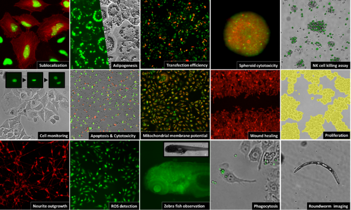
Image Credit: Scintica Instrumentation Inc.
The celloger series
Curiosis has created a new live cell imaging line. It provides researchers with advanced features, including excellent image quality and unparalleled convenience. The Celloger apparatus is small and may fit within a regular cell culture incubator (Figure 1).
Researchers may remotely view cells in real time by inserting the gadget in the incubator and connecting it to an external PC. The time-lapse tool allows you to take cell photos according to a preset schedule, which can then be readily transformed into time-lapse videos.
Using the Celloger series, clear bright-field images may be obtained utilizing contrast-enhanced optics and fluorescence images of live cells in real-time with minimal light intensity by optimizing the fluorescence filter and light route.
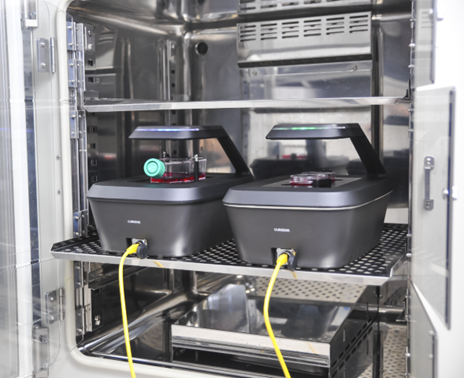
Figure 1. Celloger Mini-Plus (2 units) inside CO2 incubator. Image Credit: Scintica Instrumentation Inc.
Cytotoxicity assay
Commercially available staining reagents quantify the degree of cell death by observing phenomena in which the integrity of the cell membrane is disrupted and cell permeability increases during cell death.
Dead cells were stained with green, fluorescent CellTox™ dye to assess the cytotoxicity of nocodazole. Using the Celloger, it was determined that the number of cells evaluated by fluorescence increased cell permeability due to cell death after 20 hours.
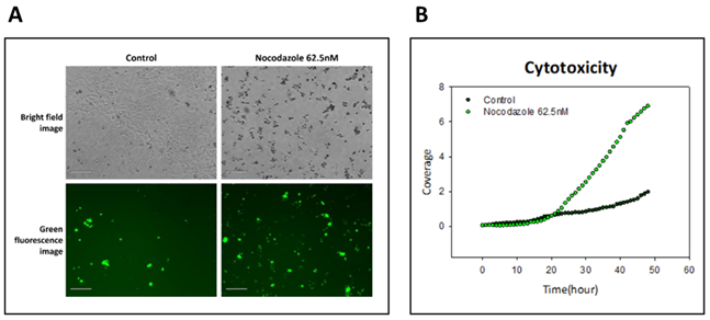
Figure 2. (A) Cell image after 35 hours of treatment with 62.5 nM nocodazole (Scale bar, 200 um). (B) Fluorescence coverage by hour. Image Credit: Scintica Instrumentation Inc.
Apoptosis assay
Apoptosis is a controlled cell death process involving membrane blebbing, cell shrinkage, and nuclear disintegration.
It was discovered that fluorescent materials were produced and detected following peptide DEVD cleavage caused by Staurosporine treatment, which is known to activate caspase and cause apoptosis (Figure 3).
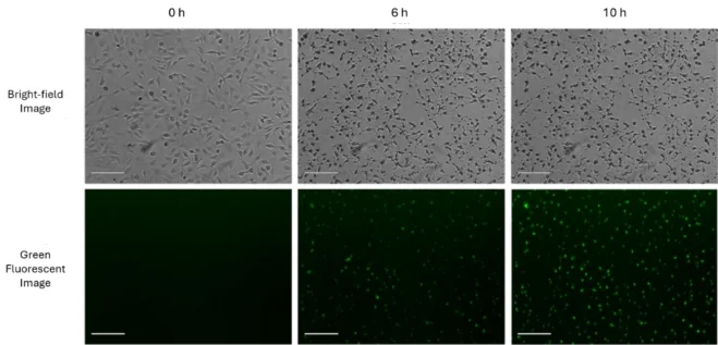
Figure 3. Using fluorescence detection of activated caspase to quantify apoptosis of Hela cells (Scale bar, 200 μm). The images were collected every 30 min using the Celloger Nano for 15 hours and 30 minutes. Image Credit: Scintica Instrumentation Inc.
Transfection assay
Transfection is a recent and effective method for inserting foreign nucleic acids into eukaryotic cells. Real-time cell imaging is useful in various applications, including assessing cell transfection effectiveness and monitoring the effects of transfected genes.
The fluorescence caused by the expression of green fluorescence protein in pCMV-GFP vector transfected in a cell was observed every 2 hours using Celloger Nano, and it was confirmed that the green fluorescence protein began to be expressed 4 hours after transfection and was maintained until 16 hours after transfection (Figure 4).
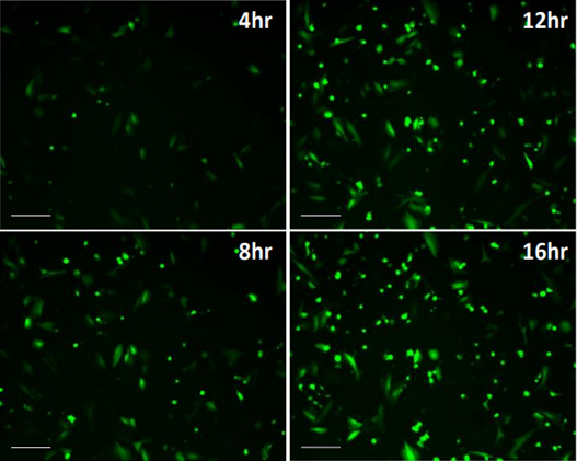
Figure 4. Time-lapse image of EGFP expression following pCMV GFP plasmid transfection (Scale bar, 200 μm). The images were collected every 2 hr for 40 hr. Image Credit: Scintica Instrumentation Inc.
Cell monitoring
Cell morphology changes occur at every major stage of the cell cycle, and it is critical to monitor these changes in cell appearance in real-time.
By monitoring cell shape (Figure 5) in real-time, researchers can discover contamination at an earlier stage, the senescence stage of the cells, and determine the ideal moment for subculture or harvesting.
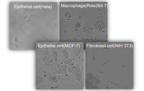
Figure 4. Time-lapse image of EGFP expression following pCMV GFP plasmid transfection (Scale bar, 200 μm). The images were collected every 2 hr for 40 hr. Image Credit: Scintica Instrumentation Inc.
Wound healing assay
The wound healing assay is the simplest and quickest approach to evaluate cell migration. When a scratch or space forms in the monolayer of cells, they demonstrate the movement process to fill in the wound until it is completely healed with new healthy cells.
Celloger’s time-lapse imaging allows for easy and effective analysis of wound healing events (Figure 6).

Figure 6. Wound healing image of HeLa cells. Image Credit: Scintica Instrumentation Inc.
Cell proliferation
Cell proliferation is the quantification of an increasing number of cells over time to ensure that the cells are growing normally.
Quantification is based on the number of fluorescently stained cells or cell confluency. In other words, a graph showing how cell number or confluency changes over time is primarily used to determine the outcome of proliferation.
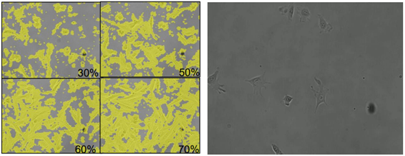
Figure 7. Cell proliferation (NIH/3T3 cell). Image Credit: Scintica Instrumentation Inc.
Real-time monitoring of spheroid cytotoxicity
2D cultures are popular due to their cost-effectiveness and ease. Nonetheless, the constraints of 2D culture methods, such as the absence of cell-to-cell or cell-to-matrix interactions and tissue-specific structures, limit their ability to simulate in vivo settings, particularly in disease models such as cancer.
Due to these constraints, there is an increasing interest in 3D culture methods that accurately depict a complex in vivo environment.
The study described below demonstrates the ability of Celloger Pro, a cutting-edge live cell imaging system, to investigate the effects of an anticancer medication, Staurosporine, on 3D spheroids derived from HEK293-GFP stable cells.
This article demonstrates the ability to dynamically capture and quantify cellular responses to drug treatment in a three-dimensional setting.
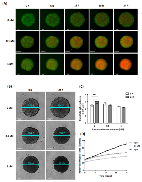
Figure 8. The results of spheroid cells with 0, 0.1, and 1 μM of SSP. (A) Merged of green and red fluorescence images for each concentration of SSP (scale bar: 200 μm). (B) Brightfield images with spheroid’s diameter (scale bar: 200 μm). (C) Comparative graph of spheroid area at 0 and 24 hours for each concentration of SSP. n=3 for each group. ***P < 0.0001 (D) Relative red fluorescence intensity graph over time. Image Credit: Scintica Instrumentation Inc.
References
- Jordan, M.A., Thrower, D. and Wilson, L. (1992). Effects of vinblastine, podophyllotoxin and nocodazole on mitotic spindles. Implications for the role of microtubule dynamics in mitosis. Journal of Cell Science, 102(3), pp.401–416. https://doi.org/10.1242/jcs.102.3.401.
- Blajeski, A.L., et al. (2002). G1 and G2 cell-cycle arrest following microtubule depolymerization in human breast cancer cells. Journal of Clinical Investigation, 110(1), pp.91–99. https://doi.org/10.1172/jci13275
- Fang, Y. and Eglen, R.M. (2017). Three-Dimensional Cell Cultures in Drug Discovery and Development. SLAS DISCOVERY: Advancing Life Sciences R&D, 22(5), pp.456–472. https://doi.org/10.1177/1087057117696795.
- Ravi, M., et al. (2014). 3D Cell Culture Systems: Advantages and Applications. Journal of Cellular Physiology, 230(1), pp.16–26. https://doi.org/10.1002/jcp.24683.
About Scintica Instrumentation Inc.
Scintica Instrumentation Inc., a high value distributor of scientific medical equipment, was created as a joint venture between two companies, Indus Instruments and ONS Projects Inc., both with long standing experience in the medical device instrumentation field. Indus Instruments is an engineering and manufacturing company with excellence in designing and producing sophisticated products for both medical and other high-tech clients in aerospace, chemical and oil and gas industries. ONS Projects Inc. is a life science investment and marketing company built on the foundation of two other successful manufacturing companies in the laboratory instrumentation field,
The principals of the two companies each have more than 25 years of experience of manufacturing, selling and supporting scientists in their research around the world. Our team consists of scientists, applications experts, engineers and sales professionals from a cross section of backgrounds, who excel at simplifying transactions and ensuring that scientists have the best equipment for achieving research excellence.
At Scintica Instrumentation, we distribute for selected manufacturers from all over the world and represent them in multiple countries including the United States, Canada, and Europe, as well as in Asia through a network of authorized sub-distributors.
Sponsored Content Policy: News-Medical.net publishes articles and related content that may be derived from sources where we have existing commercial relationships, provided such content adds value to the core editorial ethos of News-Medical.Net which is to educate and inform site visitors interested in medical research, science, medical devices and treatments.