Intravital brain imaging is a valuable tool for studying neurons, glial cells, and their surroundings in living animals. It allows the investigation of neurological illnesses, therapeutic efficacy, and the immune system. Two-photon microscopy is an invaluable tool for in vivo imaging.
It uses two lower-energy photons to stimulate fluorophores, allowing for deep-tissue imaging due to less scattering of longer-wavelength infrared light. This produces clear photos with less background noise from out-of-focus light.
Tuning two-photon lasers improves imaging by altering excitation wavelengths, allowing for more adaptable and thorough imaging across many biological situations.
The cranial imaging window (CIW) was traditionally used for brain imaging. This approach involves drilling a hole in the skull and covering the exposed brain tissue with a clear window to allow for microscopic examination.
In vivo brain imaging, however, has focused chiefly on cortical regions, posing considerable problems in detecting deeper brain structures such as the hippocampus. These constraints limit our ability to fully examine the intricate dynamics of deeper brain areas in living creatures.
The cranial imaging cannula (CIC) is a revolutionary surgical procedure that allows for real-time imaging of deep brain regions such as the hippocampus and hypothalamus. These regions are important for investigating neurodegenerative illnesses and neural dynamics.
By carefully introducing a cannula into the brain, the CIC enables long-term observation of deep brain areas with little tissue injury, making it suitable for longitudinal studies. This technique can monitor drug effects or illness progression over longer periods, such as weeks or months.
This study used tunable two-photon microscopy and the CIW approach to detect neural activity in Thy1-GCaMP6f transgenic mice. GCaMP6f is the most rapid calcium indicator for detecting cytoplasmic free calcium.
In these mice, GCaMP6f is expressed under the Thy1 promoter, allowing for the detection of neural activity via calcium changes that correspond to firing. This approach enabled us to observe GCaMP6f fluorescence in cortical neurons.
The study also used a fluorophore-conjugated CD31 antibody (CD31-Setau647) for in vivo blood vessel labeling. Using the adjustable two-photon laser, the excitation wavelength was tuned to observe both GCaMP6f and Setau647 signals.
It also investigated the depth of brain visualization possible with the CIW methodology combined with two-photon microscopy and compared it to the CIC method for accessing deeper brain regions. This comparison enabled the evaluation of the efficacy of each technique, particularly for visualizing deep brain locations.
Materials and methods
Mouse models
Thy1-GCaMP6f transgenic mice were used to track neuronal activity. These mice were genetically engineered to express GCaMP6f under the Thy1 promoter, producing steady and reproducible fluorescence in neurons.
Thy1-M transgenic mice were used to observe cortical neurons. C57BL/6N mice were used to image the hippocampus CA1 area.
Cranial image window
CIW technology was used to consistently image the brain cortex over lengthy periods. Mice were anesthetized using a mixture of Zoletil, Rumpun, and saline-injected intramuscularly to ensure adequate sedation and pain management.
Following anesthesia, the mice were placed in a stereotaxic frame to precisely target the cortex. A craniotomy was conducted, after which a cover glass was placed over the exposed brain and secured with optical bond and dental cement.
Cranial imaging cannula surgery
Following anesthesia, mice were placed in a stereotaxic frame, and a 3 mm diameter hole was created in the skull using the same methodology as CIW surgery to precisely target the hippocampus CA1 region.
After exposing the brain through a cranial hole, suction removed the meninges and a portion of the cortex, revealing the hippocampus. After the bleeding stopped, a cranial imaging cannula was introduced and covered by a 3 mm cover glass.
Except for the cannula, the incision area was sealed using a fast-curing glue and dental acrylic resin.
Blood vessel labeling
Fluorophore-conjugated antibodies (anti-CD31-Setau647, IVIM Technology) were injected intravenously into living animals to examine blood arteries. Blood vessel pictures were collected two hours after injection using intravital confocal and two-photon microscopy (IVM-CM3).
Two-photon imaging
After surgery, mice were kept in a cage to recover and followed for at least 2–4 weeks under inflammation management with Rimadyl injection (Carprofen, Zoetis).
The IVI Tag TM In Vivo Labeling Kit (IVIM Technology, Korea Republic) was used to check the blood vessels. Under anesthesia, a 25 μL Kit for labeling endothelial cells was administered intravenously through the tail vein.
After an hour, the mouse was placed on the stereotaxic stage with the heating function of the IVM-CM3 intravital microscope (IVIM Technology, Korea Republic).
Mice were placed in a stereotaxic frame to provide stability during imaging. Two-photon imaging was performed utilizing intravital confocal and two-photon microscopy (IVM-CM3).

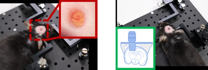
Figure 1. Mouse animal models implanted with (A) Cranial imaging window, and (B) Cranial imaging cannula. Image Credit: Scintica Instrumentation Inc.
Results

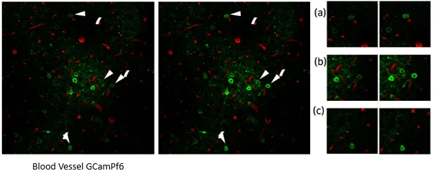
Figure 2. Visualization of GCaMP Expression. Image Credit: Scintica Instrumentation Inc.

Figure 3. Two-Photon Brain Imaging in CIW-Implanted Thy1-M-GFP Mice. Image Credit: Scintica Instrumentation Inc.
Thy1-GCaMP6f mice provide an endogenous green fluorescent protein (GFP) signal for monitoring Ca2+ ion-dependent neuronal activity.
If neurons are stimulated, GCaMP6f coupled to calcium ions quickly expresses its endogenous GFP, making it useful for investigating neuronal cell activity. This study employed the mouse species following CIW implantation to visualize real-time changes in neuronal activity.
The results (Fig 1A and 2A) revealed that some neurons had increased GFP, indicating that real-time cellular dynamics could be captured in the brain using a CIW-implanted mouse model.
The study also evaluated brain depth in Thy1-M-GFP mice implanted with a CIW using a two-photon laser at 920 nm. Second harmonic generation signals from collagen suggested it was present in the surface layers.
Blood vessels were found beneath the collagen layer, with bigger vessels in the higher sections and micro-vessels becoming more prevalent at greater depths.
Using two-photon microscopy and CIW technology, neurons labeled with Thy1-M-GFP could be visualized up to 600 μm in the cortex.

Figure 4. Two-Photon Hippocampal CA1 Imaging in CIC-Implanted Mice. Image Credit: Scintica Instrumentation Inc.
The researchers implanted a CIC into mice to overcome constraints in imaging depth and see deep brain regions. Using a 920 nm two-photon laser, they successfully identified blood vessels in the hippocampus CA1 region of CIC-implanted animals.
These findings reveal that the CIC technique enables real-time imaging of deep brain locations, such as the hippocampus, in living mice.
This innovation allows the visualization of not only cortical neuronal activity and blood vessels but also deeper brain regions such as the hippocampus, making it an invaluable tool for researching neurological illnesses and treatment responses in previously inaccessible brain locations.
References
- Dana, H., et al. (2014). Thy1-GCaMP6 Transgenic Mice for Neuronal Population Imaging In Vivo. PLoS ONE, 9(9), p.e108697. https://doi.org/10.1371/journal.pone.0108697.
- Bradley, J.E., Ramirez, G. and Hagood, J.S. (2009). Roles and regulation of Thy-1, a context-dependent modulator of cell phenotype. BioFactors, [online] 35(3), pp.258–265. https://doi.org/10.1002/biof.41.
- Benninger, R.K.P. and Piston, D.W. (2013). Two-Photon Excitation Microscopy for the Study of Living Cells and Tissues. Current Protocols in Cell Biology, 59(1), pp.4.11.1–4.11.24. https://doi.org/10.1002/0471143030.cb0411s59.
About Scintica Instrumentation Inc.
Scintica Instrumentation Inc., a high value distributor of scientific medical equipment, was created as a joint venture between two companies, Indus Instruments and ONS Projects Inc., both with long standing experience in the medical device instrumentation field. Indus Instruments is an engineering and manufacturing company with excellence in designing and producing sophisticated products for both medical and other high-tech clients in aerospace, chemical and oil and gas industries. ONS Projects Inc. is a life science investment and marketing company built on the foundation of two other successful manufacturing companies in the laboratory instrumentation field,
The principals of the two companies each have more than 25 years of experience of manufacturing, selling and supporting scientists in their research around the world. Our team consists of scientists, applications experts, engineers and sales professionals from a cross section of backgrounds, who excel at simplifying transactions and ensuring that scientists have the best equipment for achieving research excellence.
At Scintica Instrumentation, we distribute for selected manufacturers from all over the world and represent them in multiple countries including the United States, Canada, and Europe, as well as in Asia through a network of authorized sub-distributors.
Sponsored Content Policy: News-Medical.net publishes articles and related content that may be derived from sources where we have existing commercial relationships, provided such content adds value to the core editorial ethos of News-Medical.Net which is to educate and inform site visitors interested in medical research, science, medical devices and treatments.