According to the National Cancer Institute at the NIH, roughly 40.5 % of men and women will be diagnosed with cancer at some point in their lives.

γ-eye. Image Credit: Scintica Instrumentation Inc.
Depending on the type and stage of cancer, chemotherapy is a popular therapeutic option for cancer patients. Chemotherapy targets cells that grow quickly, such as cancer cells, but it can also harm normal, rapidly growing cells, such as hair follicles.
Reconfiguring the chemical structure and selectivity of existing chemotherapy medications allows for the targeted killing of cancer cells.
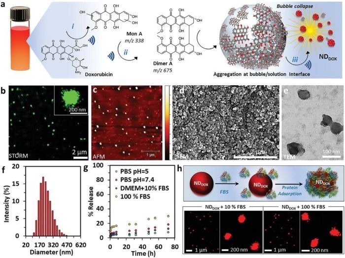
Using the traditional anticancer medicine Doxorubicin and ultrasound, researchers were able to change the structure of Doxorubicin to develop a nano drug that specifically targets and eliminates different cancer cells.
The nanodrug’s selectivity is due to its strong contact with cancer cells’ mitochondria. This results in high quantities of reactive oxygen species, which causes cell death.
In vivo biodistribution investigations revealed that the nanodrug accumulated in the lungs with negligible accumulation in the liver and spleen. In addition, the nanodrug caused low damage to healthy cells, such as fibroblasts, while killing cancer cells, including those resistant to Doxorubicin treatment.
As a result, the nano drug’s selectivity for cancer cells appears to be increased. This study demonstrates the potential of converting anticancer medications into nano drugs as a possible platform for selectively killing cancer cells.

Figure 1. Engineering DOX into a stable nanodrug. a) Schematic of the ultrasound-assisted engineering of DOX into NDDOX. A solution of DOX in water (0.5 mg mL−1) was sonicated at high frequency (490 kHz) and 2 W cm−2 for 3 h to readily form NDDOX. DOX is converted into hydroxylated species (MonA and DimA) (i) at the transient cavitation bubble/solution interface (ii) and subsequently the products self-assemble upon bubble collapse to form uniform DDOX nanoparticles (iii). b) Representative STORM image of NDDOX labeled with the photoswitchable dye AF 647; the inset shows a magnified view of a single NDDOX nanoparticle. The results are from three independent experiments. c) AFM (height 0–10 nm), d) SEM, and e) TEM images of NDDOX nanoparticles, respectively. f) The size distribution of NDDOX in aqueous solution, determined using DLS. g) Dissolution kinetics of NDDOX at pH 5 and pH 7.4 in 100 × 10−3 m PBS, 100% FBS, and cell culture medium Dulbecco’s modified Eagle medium containing 10% FBS. The % release was determined by measuring fluorescence emission (λ520 nm) of the collected supernatant after centrifugation of NDDOX. h) Scheme showing the adsorption of protein on the surface of NDDOX after incubation with FBS and STORM images of the NDDOX acquire after 8 h incubation with 10% and 100% FBS. Results are from three independent experiments. Image Credit: Scintica Instrumentation Inc.
About Scintica Instrumentation Inc.
Scintica Instrumentation Inc., a high value distributor of scientific medical equipment, was created as a joint venture between two companies, Indus Instruments and ONS Projects Inc., both with long standing experience in the medical device instrumentation field. Indus Instruments is an engineering and manufacturing company with excellence in designing and producing sophisticated products for both medical and other high-tech clients in aerospace, chemical and oil and gas industries. ONS Projects Inc. is a life science investment and marketing company built on the foundation of two other successful manufacturing companies in the laboratory instrumentation field,
The principals of the two companies each have more than 25 years of experience of manufacturing, selling and supporting scientists in their research around the world. Our team consists of scientists, applications experts, engineers and sales professionals from a cross section of backgrounds, who excel at simplifying transactions and ensuring that scientists have the best equipment for achieving research excellence.
At Scintica Instrumentation, we distribute for selected manufacturers from all over the world and represent them in multiple countries including the United States, Canada, and Europe, as well as in Asia through a network of authorized sub-distributors.
Sponsored Content Policy: News-Medical.net publishes articles and related content that may be derived from sources where we have existing commercial relationships, provided such content adds value to the core editorial ethos of News-Medical.Net which is to educate and inform site visitors interested in medical research, science, medical devices and treatments.