Interest in cell therapy has increased in recent years, with additional focus on leveraging this as a strategy to fight tumors by harnessing the immune system.
Cell therapy technologies include reprogramming patients’ own T cells to produce personalized medicines. T-cell therapy capitalizes on the immune system’s capacity to recognize and kill tumor cells- an ability that may be overwhelmed or lost as cancer develops.
Extracting and manipulating T-cells from a patient enables potential restoration of the patient’s ability to attack cancer cells. Rapid, accurate assays to qualify engineered T-cells’ potency against the target tumor cell are critical.
These assays are key to supporting the effective and efficient development of therapeutics.
This article examines a new picodroplet approach specifically designed for the functional validation of CAR-T cells. This robust and user-friendly method combines the advantages of a granzyme B assay suitable for studying cell-mediated cytotoxicity with Fluidic Sciences and Sphere Bio’s advanced high-throughput picodroplet technology (Figure 1).
This method sees engineered T cells and target cells co-encapsulated in picoliter-volume aqueous droplets (picodroplets) in an oil emulsion. A granzyme B substrate is also included.
Granzyme B can be understood as a readout of the cells’ ability to recognize and kill cancer cells. This can be captured as it is released by the T cell, enabling determination of the level of potential anti-cancer activity. This allows the rapid acquisition of cell product efficacy readouts, enabling streamlined, quality-controlled release of cell product.
An introduction to picodroplet technology
Picodroplet-based technology is a highly promising microfluidic-based technique designed for single-cell functional analysis.
This technology offers a range of advantages over traditional tools. For example, picodroplets provide a distinct microenvironment suitable for high-throughput cell-cell interaction studies and cell secretion analysis at the single-cell level.
The use of miniaturized picoliter volumes enables rapid mixing and minimal sample dilution, factors key to increasing detection sensitivity while reducing reaction time and sample requirements.
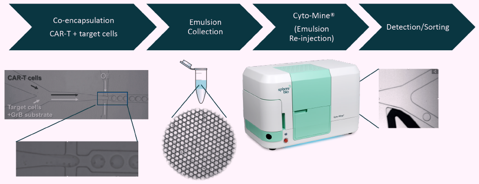
Figure 1. Workflow depicting high throughput CAR-T cell function verification in microfluidic picodroplets. Image Credit: Fluidic Sciences and Sphere Bio
Granzyme B detection in picodroplets
A commercially available granzyme B assay kit (SensoLyte® Granzyme B Activity Assay Kit, Anaspec, Fremont, CA) was adapted for use in picodroplets, allowing it to be used to detect the release of granzyme B in picodroplets.
The assay cleaves a granzyme B substrate peptide and labeling this with a 5-FAM fluorophore and a QXL®-520 fluorophore quencher. No fluorescence is emitted upon excitation of the fluorophore while it is in its intact, uncleaved state.
This is due to the nearby QXL®-520 molecule’s quenching effect (Figure 2A). The peptide substrate is cleaved in the presence of granzyme B, however, releasing the quenching molecule and prompting a fluorescent signal to be released (Figure 2A and Figure 2B).
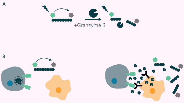
Figure 2. Principle of Granzyme B detection assay. A) Granzyme B substrate peptide labeled with 5-FAM (green star) and a QXL®-520 fluorophore quencher (grey star). No fluorescence is detected on excitation due to Fluorescence Resonance Energy Transfer (FRET) from 5-FAM to QXL®-520 (left side). On cleavage with Granzyme B the quenching molecule is removed, and fluorescence is emitted from 5-FAM (right side). B) Application of Granzyme B activity assay to CAR-T cell/target cell interaction. Left: CAR-T cell and nontarget cell, no CAR mediated signal, no Granzyme B release, 5-FAM fluorescence quenched. Right: CAR-T cell interaction with target cell results in CAR-mediated signal, Granzyme B release, substrate peptide cleavage, release of the quencher, and fluorescence, which are then detected. Image Credit: Fluidic Sciences and Sphere Bio
Granzyme B detection in picodroplets
Two populations of picodroplets at approximately 450 pL volume were initially generated using the Pico-Capture® instrument to demonstrate the assay’s general feasibility.
One of these populations contained just the fluorogenic granzyme B substrate peptide, forming a negative control, while the other population co-encapsulated recombinant granzyme B along with the substrate peptide.
Figure 3 features micrographs of both populations in brightfield (left) and green fluorescence (right) following 2 hours’ incubation at 37 ºC. It can be observed that only picodroplets containing both granzyme B and the substrate peptide (bottom) exhibit clearly detectable fluorescence after the 2-hour incubation period. In contrast, picodroplets with substrate peptide only (top) exhibit very low to no background fluorescence.
Granzyme B detection with T cells and target cells
Next, co-encapsulated polyclonal human donor T cells were genetically modified to express a CAR directed against Prostate-Specific Membrane Antigen (PSMA). This involved target cells expressing PSMA (PC3-LN3-PSMA) and the fluorogenic granzyme B substrate peptide.
An in-house custom-made biochip was employed, featuring two separate aqueous inlets to ensure that CAR-T and target cells were kept separate until immediately prior to encapsulation.
Cell concentrations were adjusted, with approximately 20% of picodroplets containing a CAR-T cell, and more than 75% of these receive at least one target cell (according to a Poisson distribution).
It is important to note that 150,000 out of 1 million generated picodroplets contained one CAR-T cell and at least one target cell, with around 15% (0.75 x 0.2) of picodroplets containing at least one CAR-T and one target cell.
CAR-T cells with PC3-LN3 cells lacking PSMA expression were also co-encapsulated as a control. This was done alongside the fluorogenic peptide substrate under otherwise identical conditions.
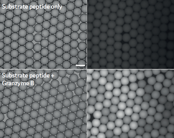
Figure 3. Detection of Granzyme B activity in picodroplets using a fluorogenic 5-FAM/ QXL®-520 substrate peptide. Brightfield (left side) and Fluorescence micrographs (right side) 2 h post generation. Scale bar: 100 μm. Image Credit: Fluidic Sciences and Sphere Bio
An Eppendorf tube was used to collect picodropets, and these were imaged under a fluorescence microscope after 2, 4, and 24 hours of incubation to detect granzyme B activity. Figure 4 features fluorescent micrographs of picodroplets containing non-expressing control cells (top) and PC3-LN3-PSMA target cells (bottom), as these were imaged after 2, 4, and 24 hours (left to right).
Picodroplets containing PC3-LN3 control cells were seen to demonstrate very little granzyme B activity after 2-4 hours. Multiple fluorescent picodroplets are indicative of granzyme B released by CAR-T cells in response to encountering one or more target cell(s) in the case of picodroplets with PC3-LN3-PSMA cells.
Increased fluorescence was observed in the negative control following prolonged incubation (24 hours), potentially due to granzyme B leaking from dead or apoptotic CAR-T cells or from other unspecific stimulation.
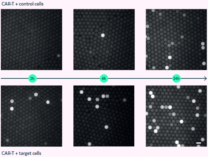
Figure 4. Granzyme B assay in picodroplets with co-encapsulated CAR-T and target cells (bottom) or control cells (top). Fluorescence micrographs of picodroplets after 2, 4, and 24 h (left to right). Scale bar: 100 μm. Image Credit: Fluidic Sciences and Sphere Bio
Granzyme B detection using Cyto-Mine®
Once it had been established that it was possible to detect granzyme B-released CAR-T cells using the 5-FAM/QXL®-520 fluorogenic peptide substrate, it was necessary to detect and sort granzyme B-positive picodroplets.
This was done using Fluidic Sciences and Sphere Bio’s Cyto-Mine® Single Cell Analysis System. CAR-T cells were co-encapsulated with PC3-LN3-PSMA target cells and the 5-FAM/QXL®-520 peptide substrate in 450 pL-sized picodroplets. This was done using Fluidic Sciences and Sphere Bio’s Pico-Capture® instrument.
The resulting emulsion was collected and loaded into a Cyto-Cartridge® to enable injection, analysis, and sorting via the Cyto-Mine® platform.
It was possible to detect the fluorescence signal immediately following the injection of all picodroplets into the Cyto-Mine® (t=0), as well as after an additional incubation of 1 hour and 2 hours at 37 ºC inside the instrument.
Figure 5 features scatter plots of picodroplet size (y-axis) versus green fluorescence (x-axis) obtained at the specified time points.
A clear fluorescent signal was detectable in around 1.25% of picodroplets at the earliest time point, with this positive population increasing over time to 2.95% of picodroplets after a 2-hour period.
Because just 15% of picodroplets contain both a CAR-T cell and at least one target cell, this figure corresponds to an actual positive rate of around 20%.
A total of 5,000 positive picodroplets were then sorted to visually confirm the presence of a fluorescent signal. The Cyto-Cartridge® was carefully removed from the instrument after sorting, and this was placed under a fluorescence microscope in order to image the picodroplets in the dispensing chamber.
Figure 6 features both a brightfield (left) and fluorescent micrograph (right) of picodroplets in the Cyto-Cartridge®’s dispensing chamber. A clear enrichment of fluorescent picodroplets is apparent.
Images were analyzed using the ImageJ software and a Hough Circle Transform algorithm, resulting in 1,112 picodroplets. It was determined that 170 of these picodroplets contained little to no fluorescence (for example, a false positive, or based on near background signal and a dark picodroplet border), while 942 picodroplets (around 85%) were deemed to be true positives.
This result corresponds to a 28-fold enrichment versus the non-sorted population with 2.95% positives after 2 hours of incubation.
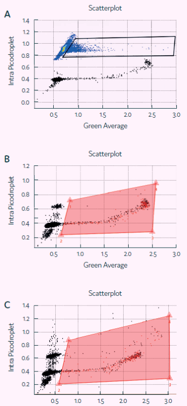
Figure 5. Detection of Granzyme B activity in picodroplets with Cyto-Mine®. Screenshots of Cyto-Mine® software during detection/ sorting, showing scatterplot of picodroplet size (Intra picodroplet, ms) against Fluorescence signal (Green Average, V) at t=0 (A), 1 h (B) and 2 h (C). The middle and bottom panel also show polygon gates used for determining percentage of positive picodroplets and for sorting. Image Credit: Fluidic Sciences and Sphere Bio
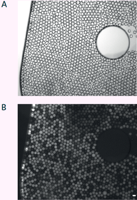
Figure 6. Brightfield (A) and fluorescent micrograph (B) of sorted picodroplets in dispensing chamber. Scale bar 100 μm. Image Credit: Fluidic Sciences and Sphere Bio
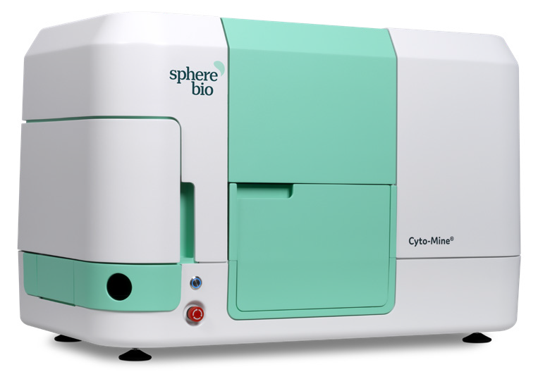
Figure 7. Cyto-Mine® - Fluidic Sciences and Sphere Bio’s automated and fully integrated single-cell analysis system. Image Credit: Fluidic Sciences and Sphere Bio
Conclusions
The study presented here demonstrated the viability of picodroplet technologies in the monitoring of CAR-T activation at relevant early timepoints following co-culture and that established assays can be successfully adapted to the picodroplet format.
Current strategies depend on screening in bulk cultures to establish CAR-T cell product profiles. Cytotoxicity readouts can be time-consuming and are not currently included in all cell products’ release criteria. This data is gathered after the fact or not at all.
The capacity to acquire a readout of a cell product’s potential efficacy fitness via a short assay is anticipated to provide significant value to cell therapy development.
Picodroplet technology enables rapid, high-throughput screening at the single-cell level, and linking this utility to granzyme B release through the co-encapsulation of CAR-T and target cells enables the development of highly functional products with the best chance of impact for patients.
Cell therapy developers could also leverage this co-encapsulation approach to interrogate other readouts of cellular fitness and cytotoxicity. These could include direct cytotoxicity of the target cells or other proteomic markers of cytotoxicity.
Bulk T cell cultures exhibit a range of phenotypic and metabolic subsets. Investigating cytotoxic efficacy at the single T cell level could provide useful data on each of these subsets individually, guiding T cell engineering towards more potent subsets.
Co-encapsulation in picodroplets could also enable the rapid screening of candidate CAR/TCR gene-modified cells for drug development.
Acknowledgments
Produced from materials originally authored by Fluidic Sciences and Sphere Bio Ltd.
About Fluidic Sciences and Sphere Bio
Fluidic Sciences develops transformative in‑solution technologies for protein interaction analysis. Its flagship Fluidity One‑M instrument leverages Microfluidic Diffusional Sizing (MDS) to measure binding affinity, stoichiometry, size, and concentration without immobilization - directly in complex backgrounds such as serum, plasma, and lysate.
Sphere Bio is a brand of Fluidic Sciences. Its technology develops and manufactures single‑cell analysis and monoclonality assurance systems that enable researchers to find, analyze, and isolate the most valuable cells with speed and precision. Its proprietary picodroplet microfluidics and Cyto‑Mine® Chroma multiplexing platform power applications across antibody discovery, cell line development, cell engineering, and cell therapy.
Sponsored Content Policy: News-Medical.net publishes articles and related content that may be derived from sources where we have existing commercial relationships, provided such content adds value to the core editorial ethos of News-Medical.Net which is to educate and inform site visitors interested in medical research, science, medical devices and treatments.