Sponsored Content by SartoriusReviewed by Maria OsipovaOct 2 2024
Until recently, the analysis of cell images was conducted manually often requiring hours of effort. Jasmine Trigg, a scientist at Sartorius, remembers this well.
“Getting the insights I needed was challenging because accurately segmenting cells or analyzing them on a large scale were frequent pain points. Another difficulty was using imaging reagents, especially with neural cells, as they were sometimes incompatible with sensitive cells or introduced artifacts,” she says, recalling her days in a neuroscience lab.
Cell data forms a critical pillar in advancing our collective knowledge of human health and finding effective treatments for diseases.
At the center of this idea is monitoring how cells grow and behave. While low-throughput or cell-destructive techniques have proven beneficial, more laboratories are incorporating non-invasive techniques to monitor live cells in real-time.
Image analysis techniques such as noise reduction, segmentation, and feature extraction are frequently deployed in such analysis. When working with manual workflows, these methodologies demand specific expertise and are vulnerable to human error. For instance, cell segmentation is a key step for identifying and separating different regions or objects within an image.
Manual segmentation requires setting intensity thresholds to separate the foreground from the background and then, with the appropriate computer software, sketching boundaries around cells or structures of interest using a computer mouse. Unsurprisingly, performing this task reproducibly and without bias on hundreds of thousands of cells is extremely difficult.
As artificial intelligence (AI) tools become commonplace in laboratories, it is hard to imagine returning to the time before they were used. For cell analysis, AI-enabled software, like the ones developed by Trigg and her team at Sartorius, has had a significant impact, automating workflows and removing subjectivity from the process.
AI-powered tools for cell analysis
The introduction of AI-driven tools has made cell culture analysis more efficient by automating segmentation, feature extraction, and data analysis. This technological advancement improves accuracy and reproducibility, allowing researchers to focus on result interpretation over conducting laborious manual tasks.
These tools utilize training data to develop models that analyze and classify cells in microscopy images with great accuracy. The training data comprises millions of high-quality images of manually annotated cells to highlight and distinguish features such as live versus dead cells or specific cell types.
Trigg is well-acquainted with this process after developing advanced AI tools for automated analysis on the Incucyte® Live-Cell Analysis System.
“One thing you realize is how crucial it is to have high-quality ground truth data for training and validating the models. First, you need to be clear about what you want the model to do and the contexts in which it will be used. Second, it is important to be specific and consistent when annotating images for model training, and to include a wide range of examples. Finally, you must have good communication and understanding between software engineers and biologists to ensure the model works as intended.”
The team used this process to develop the Incucyte® AI Cell Health Analysis Software Module, one of the new AI tools that can be used with the Incucyte® system. Using this tool, scientists can accurately process and quantify live or dead cells without fluorescent dyes, enhancing the biological relevance of their data.
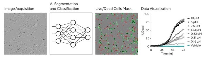
Image Credit: Sartorius
AI cell health analysis applications
Live/dead analysis is a crucial part of evaluating overall cell health. It is critical for assessing cell viability and understanding cellular responses to treatments or conditions.
This process can be automated using the Incucyte® AI Cell Health Analysis Module—it can kinetically quantify live and dead adherent and non-adherent cells over time, making it a valuable tool in cell culture studies.
As Trigg clarifies, “AI tools provide unbiased cell analysis through a simple process, leading to better insights from both complex images and straightforward assays. They can accurately segment cells without interference from background elements like texture, scratches, or precipitation, and allow consistency across multiple conditions, significantly reducing time spent on analysis, especially at higher throughputs.”
This module is used across numerous research fields, especially those that require real-time monitoring and cell health analysis.
Cancer research
The module is used in cancer research to evaluate drug cytotoxicity and investigate the effects of several therapeutic compounds on tumor cells. Researchers take advantage of its capabilities to run drug sensitivity assays, allowing for the quantification of live and dead cells without fluorescent labeling, which is key for examining primary cells and tumor cell lines.
Neuroscience
One of the challenges of working with neural cell types is the complexity of their morphology and behavior, which makes manual analysis strenuous and error-prone. The Incucyte® AI Cell Health Analysis Module is a key part of segmenting these intricate morphologies accurately to support an understanding of neuronal health and the impact of various treatments on glial cell health.
Drug development
AI-based image analysis is central to high-throughput screening processes in drug development. It enables streamlined compound testing across numerous cell types and conditions. Its capability to deliver real-time, label-free analysis improves the throughput and reliability of drug-testing protocols.
Conclusion
Machine learning techniques, particularly convolutional neural networks, provide valuable opportunities to streamline preprocessing and feature extraction, significantly improving image quality and accuracy in routine cell analysis.
Sartorius is at the forefront of providing software solutions like the AI Cell Health Module and the AI Confluence Module. These tools improve the precision and efficiency of live-cell analysis, facilitate non-invasive, image-based measurements of cell growth, and promote an advanced understanding of complex cellular processes.
About Sartorius

Sartorius is a leading international pharmaceutical and laboratory equipment supplier. With our innovative products and services, we are helping our customers across the entire globe to implement their complex and quality-critical biomanufacturing and laboratory processes reliably and economically.
The Group companies are united under the roof of Sartorius AG, which is listed on the Frankfurt Stock Exchange and holds the majority stake in Sartorius Stedim Biotech S.A. Quoted on the Paris Stock Exchange, this subgroup is comprised mainly of the Bioprocess Solutions Division.
Innovative technologies enable medical progress
A growing number of medications are biopharmaceuticals. These are produced using living cells in complex, lengthy and expensive procedures. The Bioprocess Solutions Division provides the essential products and technologies to accomplish this.
In fact, Sartorius has been pioneering and setting the standards for single-use products that are currently used throughout all biopharmaceutical manufacturing processes.
Making lab life easier
Lab work is complex and demanding: Despite repetitive analytical routines, lab staff must perform each step in a highly concentrated and careful way for accurate results.
The Lab Products and Services Division helps lab personnel excel because its products, such as laboratory balances, pipettes and lab consumables, minimize human error, simplify workflows and reduce physical workloads.
Sponsored Content Policy: News-Medical.net publishes articles and related content that may be derived from sources where we have existing commercial relationships, provided such content adds value to the core editorial ethos of News-Medical.Net which is to educate and inform site visitors interested in medical research, science, medical devices and treatments.