Sponsored Content by SartoriusReviewed by Maria OsipovaDec 5 2024
Neurodegenerative diseases, like Parkinson’s Disease (PD), are debilitating chronic disorders that cause degeneration over time and/or the death of neuronal cells. Better in vitro models, which can recapitulate the chronic characteristics of the disease, combined with advanced cell models such as primary cells or iPSCs in mono- or co-culture, have the potential to improve insights into the pathology of the disease, contributing to drug discovery. However, conventional approaches used to research these dynamic biological changes present a challenge.
Research into these complex and sensitive models demands advanced technologies, like live-cell analysis, that can follow and robustly quantify dynamic cellular processes in non-perturbing conditions.
Challenges
- Studying complicated and disease-relevant neuronal models for long durations at a high-throughput.
- Methods for quantifying neuronal morphology or toxicity may be perturbing, only use a single end-point, or are incompatible with sensitive models.
- Techniques that assess phagocytosis can have destructive or laborious protocols and do not allow monitoring of the peptide engulfment in real-time.
Solutions
- Live-cell analysis enables real-time visualization and quantification of in vitro models in an environment that is physiologically relevant in up to 96/384-well microplate formats.
- The Incucyte® Neurite Outgrowth Assay is non-perturbing and offers compatibility with sensitive cell types, inclusive of iPSC-derived or primary cells, in mono- or co-culture.
- The Incucyte® Phagocytosis Assay offers an easy-mix- read approach for the kinetic visualization and quantifications of phagocytosis and assessing disease-associated peptides.
Quantitative pharmacology in PD co-culture models
Monitoring neurite dynamics in long-term in vitro cell cultures is crucial in characterizing and evaluating neurodegenerative disease models and assessing pharmacological neurotoxic effects. The Incucyte® Live-Cell Analysis System combined with the Incucyte® Neurotrack Analysis Software Module can kinetically quantify neuronal outgrowth.
This assay allows for analyzing neurons in monoculture (label-free) or in coculture with astrocytes, utilizing a non-perturbing neuron-specific Incucyte® Neurolight Orange Lentivirus, for continuous analysis of branch points and neurite length.
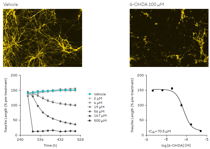
To develop a model of PD, a co-culture system of rat primary striatal neurons and astrocytes was created in a 96-well microplate (Figure 1). After infecting the neurons with neurolight orange and a session of neurite development (10 days), the dopaminergic-specific neurotoxin 6-hydroxydopamine (6-OHDA) was used to induce diseased-relevant neuronal damage. This was monitored with live-cell analysis.
A time and concentration-dependent effect on neurite disruption was noted, with a yield of an IC₅₀ value for 6-OHDA of 70.5 μM for neurite length.
This model is also useful in assessing neurotoxic compound specificity over a range of brain regions (Figure 2). To explore this further, co-cultures of rat primary striatal, substantia nigra, or cortical neurons and astrocytes expressing neurolight orange were treated with 6-OHDA after 10 days.
Bar graphs and drug-response curves demonstrate the selective profile of the drug to neurite length in a variety of brain regions, with 6-OHDA having a selective effect on the nigra and striatal neurons. The vehicle data shows the differential development of neurite formation over several brain regions.
Live-cell analysis combined with the neurite outgrowth assay allows for monitoring complex in vitro models in a non-perturbing way with the flexibility of a range of cell types, such as primary, iPSC-derived, or immortalized neurons in mono- or co-culture. This approach offers a robust high-throughput assay that detects pharmacological effects on neurite dynamics and helps screen neuroprotective agents.

Figure 1. 6-OHDA-induced Neurite Disruption in a PD Model. Concentration-dependent neurotoxic effects of 6-OHDA on neurite length measured using live-cell analysis in rat primary co-culture models. Image Credit: Sartorius

Figure 2. 6-OHDA selectively effects neurons from different brain regions. Rat primary neurons co-cultured with astrocytes respond differently to 6-OHDA, with live-cell analysis showing selective effects on substantia nigra and striatal neurite outgrowth compared to cortical regions. Image Credit: Sartorius
Phagocytosis of disease-associated peptides
Microglia are the brain’s resident macrophage and are critical in the neuro-inflammatory response. Aggregated peptides, shown to induce neuropathology, are quickly phagocytosed by microglia.
Live-cell analysis can be used to assess phagocytosis of peptides associated with PD (Figure 3). Here, over two weeks, iPSC microglia precursor cells were differentiated in a 96-well plate to mature microglia. Aggregated α-synuclein peptides were labeled with the pHrodo® Orange Cell Labeling Kit for Incucyte® and subsequently added to the iPSC-derived microglia at various concentrations. Phagocytosis was quantified through an increase in fluorescence.
The results demonstrated that iPSC-derived microglia quickly engulf aggregated α-synuclein, and uptake in a time- and peptide concentration-dependent manner was seen. The phagocytosis assay offers a simple but powerful solution for visualizing iPSC-derived microglia and the kinetic quantification of phagocytosis for disease-relevant peptides in a high-throughput format. This approach can potentially facilitate the development of new neuro-therapeutics that target microglial function.

Figure 3. Phagocytosis of α-synuclein in PD neuro-inflammatory model. iPSC-derived microglia engulf aggregated α-synuclein in a time- and concentration-dependent manner, as shown by an increase in orange fluorescence area following engulfment. Image Credit: Sartorius
Summary
This case study explores using live-cell analysis to improve the study of PD models. It showcases the challenges of conventional methods when studying complex neuronal models and offers live-cell analysis as a potential solution. This technology enables real-time visualization and quantification of cellular processes in a non-perturbing way, utilizing tools such as the Incucyte® Neurite Outgrowth and Phagocytosis Assays. These assays allow for monitoring neurite dynamics and phagocytosis in PD models, which provides insights into neurotoxicity and neurodegeneration. The approach supports high-throughput screening and development of neuroprotective agents against disease-associated toxicity and neuroinflammation.
About Sartorius

Sartorius is a leading international pharmaceutical and laboratory equipment supplier. With our innovative products and services, we are helping our customers across the entire globe to implement their complex and quality-critical biomanufacturing and laboratory processes reliably and economically.
The Group companies are united under the roof of Sartorius AG, which is listed on the Frankfurt Stock Exchange and holds the majority stake in Sartorius Stedim Biotech S.A. Quoted on the Paris Stock Exchange, this subgroup is comprised mainly of the Bioprocess Solutions Division.
Innovative technologies enable medical progress
A growing number of medications are biopharmaceuticals. These are produced using living cells in complex, lengthy and expensive procedures. The Bioprocess Solutions Division provides the essential products and technologies to accomplish this.
In fact, Sartorius has been pioneering and setting the standards for single-use products that are currently used throughout all biopharmaceutical manufacturing processes.
Making lab life easier
Lab work is complex and demanding: Despite repetitive analytical routines, lab staff must perform each step in a highly concentrated and careful way for accurate results.
The Lab Products and Services Division helps lab personnel excel because its products, such as laboratory balances, pipettes and lab consumables, minimize human error, simplify workflows and reduce physical workloads.
Sponsored Content Policy: News-Medical.net publishes articles and related content that may be derived from sources where we have existing commercial relationships, provided such content adds value to the core editorial ethos of News-Medical.Net which is to educate and inform site visitors interested in medical research, science, medical devices and treatments.