Biotin and avidin or streptavidin (SA) exhibit a very powerful and specific interaction. This makes it a perfect tool in protein biochemistry. But producing high-quality biotinylated proteins presents a significant challenge.
Scientists usually face technical problems like high batch-to-batch variation, inadequate labeling and uncertain performance. Additionally, the selection of biotinylation methods also considerably impacts the ultimate result. Every target protein (assay) exhibits its special biochemical characteristics, and thus needs a tailored solution.
These perplexing factors make it hard for laboratories to prepare and use biotin conjugates, while only a few products are available in the market. To bridge this gap, ACROBiosystems has come up with a unique collection of pre-labeled and experimentally validated biotinylated recombinant proteins. The Mabsol® biotinylated protein collection includes over a hundred generally studied drug targets and biomarker proteins.
The company makes use of high-quality recombinant proteins that are as close as possible to natural conformation and modification as starting materials. Also, they completely go through each option at hand to realize the highest detection sensitivity and bioactivity. The company ensures the most rigorous quality control measures to guarantee the least batch-to-batch variations.
Mabsol® biotinylated protein collection consists of two special and complimentary product series — the PrecisionAvi series built upon the Avitag™ technology, and the UltraLys series produced using the in-house developed chemical labeling method. These products are created with every attention to detail.
Products
PrecisionAvi series
An exclusive collection of ready-to-use Avitag™ biotinylated proteins
The products in this range are solely produced with the help of Avitag™ technology. In short, a special 15 amino acid peptide, the Avi tag, is added to the recombinant protein at the time of expression vector construction. The single lysine residue present in the Avi tag is enzymatically biotinylated by the E. Coli biotin ligase called BirA.
This single-point enzymatic labeling method offers several benefits for binding assays that find regular use.
- The biotinylation only takes place on the lysine residue of the Avi tag
- The protein orientation is even when immobilized on an avidin-coated surface
- There is no interference with the natural binding activities of the target protein
All products
Table 1. Source: ACROBiosystems
| . |
| 2B4 |
4-1BB |
4-1BB Ligand |
ACE2 |
Alpha-Synuclein |
Angiopoietin-2 |
Angiopoietin-like 3 |
| ANGPTL7 |
APRIL |
B7-1 |
B7-2 |
B7-H3 |
B7-H3 (4Ig) |
B7-H4 |
| B7-H5 |
B7-H6 |
B7-H7 |
BAFF |
BAFFR |
BCMA |
BLAME |
| BTLA |
BTN1A1 |
BTN2A1 |
BTN3A1 |
BTN3A2 |
CA125 |
Carbonic Anhydrase IX |
| CBLB |
CD133 |
CD14 |
CD155 |
CD161 |
CD19 |
CD2 |
Click here to learn about many more products.
UltraLys series
A special series of chemically labeled biotinylated proteins with ultra-sensitivity
In this series, the products are produced with the in-house developed chemical labeling method. The main amines in the side chains of lysine residues and the N-terminus of protein are conjugated with biotins, generally leading to several biotin attachments on a single protein molecule.
- Higher detection sensitivity can be achieved
- Comes with an in-house developed chemical labeling method
All products
Table 2. Source: ACROBiosystems
| . |
| Adalimumab |
B7-H4 |
Bevacizumab |
CD19 |
CD24 |
CD3
epsilon |
CD3E &
CD3D |
| Cetuximab |
CX3CL1 |
DNP |
EpCAM |
EphB4 |
ErbB3 |
FcRn
(FCGRT & B2M) |
| FMC63 |
GM-CSF |
GPA33 |
Growth
Hormone R |
Her2 |
IL-6 |
Mesothelin |
| NKp46 |
Nucleocapsid
protein |
Protein L |
SOST |
Spike RBD |
TFPI |
TNF-alpha |
| Transferrin R |
VEGF165 |
Vitronectin |
|
|
|
|
Applications
- SPR
- ELISA
- BLI
- FACS
- Enrichment
- Biopanning
Key features
Closest to natural conformation and modification
The production of recombinant proteins, like the biotinylated proteins, is performed with the help of the proprietary HEKMax® expression platform. As expression hosts, the human HEK293 cells offer a range of benefits over other cell types as outlined in the table below.
Table 3. Source: ACROBiosystems
| Expression Systems |
Folding |
Phosphorylation |
Proteolytic Processing |
Glycosylation |
| E.Coli |
+ |
N/A |
N/A |
N/A |
| Insect Cell |
++ |
++ |
++ |
Poor |
| Plant Cell |
+++ |
+++ |
+++ |
Poor |
| CHO Cell |
++++ |
++++ |
++++ |
Non-Human Like |
| Human Cell |
+++++ |
+++++ |
+++++ |
Human Authentic |
High bioactivity and detection sensitivity
The bioactivity of biotinylated proteins is identified through the structure of the protein itself and the way biotinylation is executed. For each protein, the company tests multiple options of tags and biotinylation approaches, while assessing the products in a range of binding assays. However, only those that exhibit the best performance are chosen for production. Figure 1 displays the internal evaluation experiments.
Figure 1. Binding activity of different forms of biotinylated PD1 evaluated in a functional ELISA against rhPD-L1 (Cat. No. PD1-H5258). Image Credit: ACROBiosystems
High bioactivity and excellent detection sensitivity in different applications are offered by the biotinylated proteins from ACROBiosystems. In functional ELISA, the biotinylated human TACI exhibits high binding activity with BAFF (see Figure 2).
Fascinatingly, oriented immobilization of biotinylated CD155 provides much greater sensitivity compared to random immobilization of unconjugated CD155 (see Figure 3). From Figures 4 and 5, it can be seen that the biotinylated PD1 can be used on AlphaLISA to determine the binding activity with its ligands PD-L1 and PD-L2.
Figures 6 and 7 illustrate that it is easy to use the biotinylated protein for identifying the affinity between protein and therapeutic antibody in SPR assay. Furthermore, the biotinylated proteins are usually utilized in cell-based assay, for example, cytotoxicity assay and assessment of CAR expression (see Figure 8).
ELISA
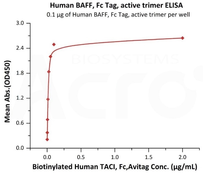
Figure 2. Immobilized Human BAFF, Fc Tag, active trimer (Cat. No. BAF-H5261) at 1 μg/mL (100 μL/well) can bind Biotinylated Human TACI, Fc, Avitag (Cat. No. TAI-H82F6) with a linear range of 0.002–0.05 μg/mL. Image Credit: ACROBiosystems
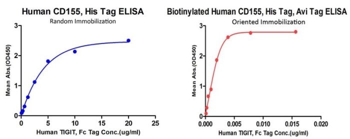
Figure 3. Comparison of oriented immobilization (biotinylated CD155 on precoated streptavidin plate, Cat. No. CD5-H82E3) and random immobilization (unconjugated CD155 direct coating on 96-well plate, Cat. No. CD5-H5223) binding to TIGIT (Cat. No. TIT-H5254). Oriented immobilization of biotinylated CD155 offers much higher sensitivity than random immobilization of unconjugated CD155. Image Credit: ACROBiosystems
AlphaLISA
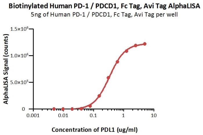
Figure 4. Biotinylated Human PD-1 (Cat. No. PD1-H82F1) at 1 μg/mL (5 μL/well) can bind Human PDL1 (Cat. No. PD1-H5229) with a linear range of 0.02–0.625 μg/mL. Image Credit: ACROBiosystems
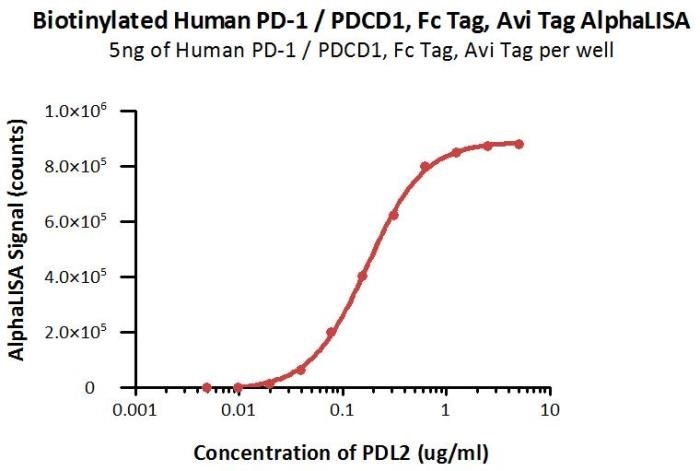
Figure 5. Biotinylated Human PD-1 (Cat. No. PD1-H82F1) at 1 μg/mL (5 μL/well) can bind Human PDL2 (Cat. No. PD2-H5220) with a linear range of 0.02–0.625 μg/mL. Image Credit: ACROBiosystems
SPR
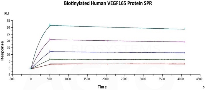
Figure 6. Immobilized biotinylated human VEGF165 (Cat. No. VE5-H82Q0) on CM5 Chip via streptavidin, can bind Avastin with an affinity constant of 0.417 nM as determined in SPR assay (Biacore T200). Image Credit: ACROBiosystems
BLI
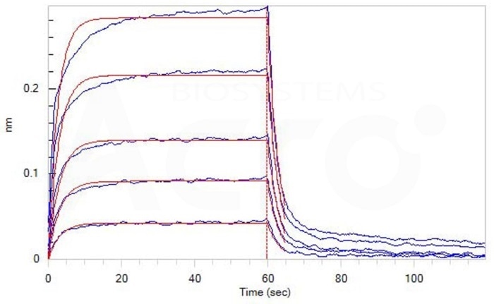
Figure 7. Loaded Biotinylated Human PD-1, Avitag, His Tag (recommended for biopanning) (Cat. No. PD1-H82E4) on SA Biosensor, can bind Human PD-L1, His Tag (HPLC verified) (Cat. No. PD1-H5229) with an affinity constant of 2.4 μM as determined in BLI assay (ForteBio Octet Red96e). Image Credit: ACROBiosystems
Cell-based assay
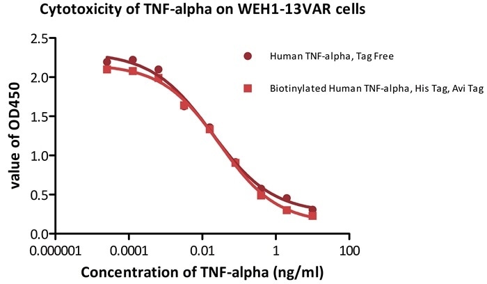
Figure 8. Recombinant human TNF-alpha (Cat. No. TNA-H82E3) induces cytotoxicity effect on the WEH1-13VAR cells in the presence of the metabolic inhibitor actinomycin D. The EC50 for this effect is 0.014–0.029 ng/mL. The result shows that the biotinylated human TNF-alpha is consistent with naked TNF-alpha in cytotoxicity assay. Image Credit: ACROBiosystems
Low batch-to-batch variation
ACROBiosystems regularly applies strict quality control measures to guarantee the consistent performance of the products. Newly introduced products undergo side-by-side comparison with the internal standard in a range of assays.
Only the products within an acceptable margin of variation are allowed to be launched. As illustrated in Figure 9, three distinct batches of biotinylated hTNF-alpha (Cat. No. TNA-H82E3) were tested and compared with the help of a standard ELISA analysis against Adalimumab. The results indicate that the batch difference among the tested samples is negligible. Also, the batch-to-batch conformance is good as shown in cell-based assay (see Figure 10).
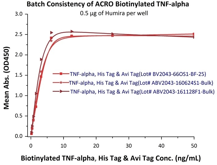
Figure 9. In the above ELISA analysis, three different lots of biotinylated hTNF-alpha (Cat. No. TNA-H82E3) were used to detect immobilized Adalimumab (0.5 μg/mL). The result showed that the batch variation among the tested samples is negligible. Image Credit: ACROBiosystems
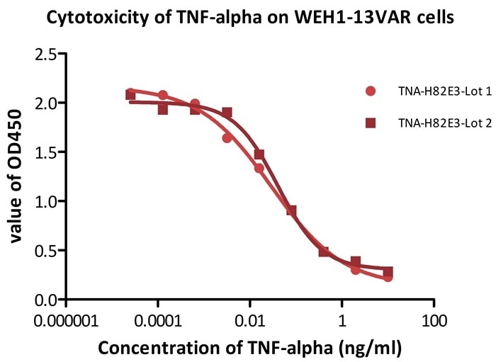
Figure 10. Recombinant biotinylated human TNF-alpha (Cat. No. TNA-H82E3) induces cytotoxicity effect on the WEH1-13VAR cells in the presence of the metabolic inhibitor actinomycin D. The EC50 for this effect is 0.029–0.052 μg/mL. Image Credit: ACROBiosystems